holotype
Nature du spécimen309 image(s) · 0 Actualités
Galerie d'images
![Figure 1: Geographic provenance and speculative reconstruction of the gigantic titanosaurian sauropod dinosaur Notocolossus gonzalezparejasi gen. et sp. nov.
(a) Type locality of Notocolossus (indicated by star) in southern-most Mendoza Province, Argentina. (b) Reconstructed skeleton and body silhouette in right lateral view, with preserved elements of the holotype (UNCUYO-LD 301) in light green and those of the referred specimen (UNCUYO-LD 302) in orange. Scale bar, 1 m. (All images were hand drawn by the senior author [B.J.G.R.] and subsequently edited using Adobe Illustrator software.)](https://upload.wikimedia.org/wikipedia/commons/thumb/7/71/Notocolossus.jpg/330px-Notocolossus.jpg)
Figure 1: Geographic provenance and speculative reconstruction of the gigantic titanosaurian sauropod dinosaur Notocolossus gonzalezparejasi gen. et sp. nov. (a) Type locality of Notocolossus (indicated by star) in southern-most Mendoza Province, Argentina. (b) Reconstructed skeleton and body silhouette in right lateral view, with preserved elements of the holotype (UNCUYO-LD 301) in light green and those of the referred specimen (UNCUYO-LD 302) in orange. Scale bar, 1 m. (All images were hand drawn by the senior author [B.J.G.R.] and subsequently edited using Adobe Illustrator software.)
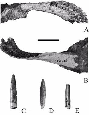

Cranial and postcranial sauropod remains from sediments of the Praia da Amoreira-Porto Novo Fm. of the coastal sector of Praia da Consolação-Lourinhã-Torres Vedras: 1-2, ?Turiasauria indet., heart-shaped tooth (SHN (JJS) 142, Praia da Corva) in lingual (1) and labial (2) views; 3-4, ?Macronaria indet., spatulate tooth (SHN 513, Porto Novo) in labial (3) and lingual (4) views; 5-6, Macronaria indet., compressed cone-chisel-shaped tooth (SHN 578, Valmitão) in lingual (5) and labial (6) views; 7-8, Eusauropoda indet., partial left maxilla (SHN 582, Praia dos Frades) in lateral (7) and posterior (8) views; 9, Titanosauriformes indet., posterior caudal vertebra (SHN 523, Praia da Corva) in right view; 10-12, Diplodocinae indet., partial skeleton (SHN (JJS) 177, Valmitão), anterior caudal neural spine in posterior view (10), anterior caudal centrum in right view (11) and left ischium in medial view (12); 13-16, holotype material of Zby atlanticus (ML 368, Vale de Pombas), right ungueal I in lateral view (13), right humerus in anterior view (14), right radius in posterior view (15), right ulna in lateral view (16); 17, Eusauropoda indet., partial distal forked-chevron (SHN 587, Praia da Corva) in medial view; 18, Sauropoda indet., pedal ungueal I (SHN 524, Praia de Pedrogãos) in lateral view; 19-22, Macronarian indet., partial skeleton (SHN 181, Valmitão), right astragalus in proximal view (19), anterior caudal vertebra in anterior view (20), right tibia in lateral view (21) and right fibula in medial view (22); 23-24, Eusauropoda indet., partial skeleton (SHN 530, Praia da Corva), anterior chevron in anterior view (23) and anterior caudal vertebra in posterior view (24); 25, cf. Duriatitan humerocristatus, humerus (MG 4976, Praia dos Frades) in anterior view; 26, Sauropoda indet., partial skeleton (SHN 534, Santa Rita), middle chevron in posterior view; 27, Diplodocidae indet., partial skeleton (SHN (JJS) 179, Praia Vermelha), dorsal/caudal (?) neural spine in posterior view; 28-30, holotype material of Dinheirosaurus lourinhanensis (ML 414, Porto Dinheiro), proximal end of a dorsal rib in anterior view (28), anterior caudal neural spine in posterior view (29) and articulated dorsal vertebrae in right view (30). Black scale bar: 10 cm; Grey scale bar: 5 cm; Brown scale bar: 1 cm. See Anatomical abbreviations for abbreviations.

(A) Blikanasaurus cromptoni (holotype SAM K403; from Galton & Van Heerden, 1985).
(A) Blikanasaurus cromptoni (holotype SAM K403; from Galton & Van Heerden, 1985).

Holotype specimen of Pterodactylus suevicus (now classified as Cycnorhamphus), specimen number GPIT 80 (example 53 of Wellnhofer 1970)
Holotype specimen of Pterodactylus suevicus (now classified as Cycnorhamphus), specimen number GPIT 80 (example 53 of Wellnhofer 1970)
Holotype specimen of Pterodactylus suevicus (now classified as Cycnorhamphus), specimen number GPIT 80 (example 53 of Wellnhofer 1970)

Cimoliopterus cuvieri. Holotype NHMUK PV 39409 (Cenomanian / Turonian, Chalk Formation), anterior part of the rostrum A right lateral view B respective line drawing C ventral view D respective line drawing. Abbreviations: m – maxillae, pm – premaxillae, pmcr – premaxillary crest, prid – palatal ridge. Arrows and numbers indicate alveoli or teeth and their respective position. Scale bar = 10 mm. Photos courtesy of The Natural History Museum.


Camposipterus nasutus comb. n. Holotype CAMSM B 54556 (Albian, Cambridge Greensand), anterior part of the rostrum A left lateral view B respective line drawing C ventral view D respective line drawing. Abbreviations: m – maxillae, pm – premaxillae, prid – palatal ridge. Arrows and numbers indicate alveoli or teeth and their respective position. Scale bar = 10 mm.

Holotype skeleton of Jianchangnathus robustus on display at the Paleozoological Museum of China.

Holotype of Ulughbegsaurus as well as the holotype placed on a reconstruction of Ulughbegsaurus's skull

Specimens of Galleonosaurus dorisae n. gen. n. sp. from the Flat Rocks Sandstone in the upper Barremian, Wonthaggi Formation, Gippsland Basin, southeastern Australia: (1–2) holotype (NMV P229196), left maxilla in lateral (1) and medial (2) views; (3) NMV P208178, left maxilla in lateral view; (4) NMV P212845, left maxilla in lateral view; (5) NMV P209977, left maxilla in lateral view; (6) NMV P186440, left maxilla in lateral view; (7) NMV 208113, right maxillary tooth in labial view. Scale bars = 10 mm (1–6); 1 mm (7).


Partial skeleton of Claosaurus agilis (holotype YPM 1190). (A) Right ilium in lateral view. (B) Partial postorbital in lateral view. (C) Distal process of the right ischium in lateral view. (D) Mounted partial skeleton of YPM 1190. (E) Coronoid process of the right dentary in lateral view. (F) Fragment of maxilla in lateral view. (G) Detail of the maxillary tooth crowns in (F). (H) Fragment of maxilla in lateral view. (I) Detail of a maxillary tooth crown in (H).