holotype
Nature du spécimen309 image(s) · 0 Actualités
Galerie d'images

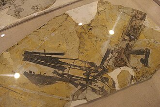
Figure description from paper: "A Cast of BES SC 999, the holotype of Besanosaurus leptorhynchus and (B) interpretative drawing (modified from Dal Sasso & Pinna, 1996). Foetal remains are highlighted with green lines. a astragalus, c calcaneum, Cl clavicle, Co coracoid, Fe femur, Fi Fibula, H humerus, i intermedium, Il Ilium, Is Ischium, P pubis, p pisiform, R radius, r radiale, S scapula, T Tibia, U Ulna, u ulnare; 2, 3, and 4, distal carpals and tarsals; II, III, IV, and V, metacarpals and metatarsals. The apostrophe (‘) indicates left elements. Scale bar represents 50 cm"
Figure description from paper: "A Cast of BES SC 999, the holotype of Besanosaurus leptorhynchus and (B) interpretative drawing (modified from Dal Sasso & Pinna, 1996). Foetal remains are highlighted with green lines. a astragalus, c calcaneum, Cl clavicle, Co coracoid, Fe femur, Fi Fibula, H humerus, i intermedium, Il Ilium, Is Ischium, P pubis, p pisiform, R radius, r radiale, S scapula, T Tibia, U Ulna, u ulnare; 2, 3, and 4, distal carpals and tarsals; II, III, IV, and V, metacarpals and metatarsals. The apostrophe (‘) indicates left elements. Scale bar represents 50 cm"

The holotype of Dianmeisaurus mutaensis (HFUT MT-21-08-001). (A) the skeleton in dorsal view; (B) the counterpart of (A) (natural mold). Scale bars equal 1 cm.

Holotype skull of Plesiotylosaurus crassidens (LACM 2759) on display at the Natural History Museum of Los Angeles County.

Austriadactylus cristatus from Carnic Prealps and illustration of holotype (C).

Holotype specimen (GMV2128) of Dendrorhynchoides curvidentatus on display at the Geological Museum of China.


T. regalis FSAC-OB 1, holotype left humerus. In (A), ventral view, (B) dorsal view, (C) anterior view, (D) distal view, and (E) proximal view. Abbreviations: dpc, deltopectoral crest; ect, ectepicondyle; ent, entepicondyle; lc, lateral condyle; mc, medial condyle; pf, pneumatic fossa/foramen; scpr, supracondylar process; uc, ulnar crest; ut, ulnar tubercle.

Eopteranodon lii holotype (BPV 078). (A) Counterpart; (B) main part. (C and D) Respective schematic drawings. Abbreviations: cv, cervical vertebra; co, coracoid; d1–d4, digits 1–4; fe, femur; fi, fibula; h, humerus; j, jugal; mand, mandible; mc, metacarpal; pmc, premaxillary crest; pe, pelvis; ph, phalanx; ti, tibia; ul, ulna; rad, radius. Scale bars: C, 50 mm; D, 10 mm.


The holotype dorsal neural arch of Saurophaganax maximus OMNH 1123 in A, anterior; B, posterior; C, left lateral; D, right lateral; and E, ventral views. Abbreviations: acdl, anterior centrodiapophyseal lamina; al, accessory lamina; cpol, cen-tropostzygapophyseal lamina; cprl, centroprezygapophyseal lamina; di, diapophysis; nc, neural canal; pa, parapophysis; pcdl, posterior centrodiapophyseal lamina; poz, postzygapophysis; prz, prezygapophysis; spol, spinopostzygapophyseal lamina; sprl?, possible spinoprezygapophyseal lamina; tpol, intrapostzygapophyseal lamina; tprl, intraprezygapophyseal lamina.

Skeletal diagram of Veterupristisaurus milneri including all known remains. The middle vertebrae is the holotype (MB R 1938), the pair on the right are the paratypes (MB R 2166), and the far left caudal is the questionably referred vertebra (MB R 1940).


Most components of the holotype specimen of Beipiaosaurus inexpectus, IVPP V 11559. Derived from Supplementary Figure 1 in the source.