←

os

Partie anatomique

76 image(s) · 55 Actualités

Galerie d'images

Mounted replica of a composite skeleton of Edmontosaurus annectens on display at the University of Oxford Museum, Oxford, England. The original skeleton is compiled from disarticulated fossil bones from a bonebed of the Hell Creek Formation, exposed in the Ruth Mason Quarry in Harding County, South Dakota. It is 8.5 m (28 ft.) long and the skull is almost 1 m (39 in.) in length.[1][2]


↑ Dinosaurs in the Museum. Oxford University Museum of Natural History (brochure, PDF), p. 7

↑ BHI Fossil Replica Catalog 2012. Black Hills Institute of Geological Research, Inc., Hill City, SD, 2012 (PDF), p. 22
Taxons Saurolophini

Mounted replica of a composite skeleton of Edmontosaurus annectens on display at the University of Oxford Museum, Oxford, England. The original skeleton is compiled from disarticulated fossil bones from a bonebed of the Hell Creek Formation, exposed in the Ruth Mason Quarry in Harding County, South Dakota. It is 8.5 m (28 ft.) long and the skull is almost 1 m (39 in.) in length.[1][2] ↑ Dinosaurs in the Museum. Oxford University Museum of Natural History (brochure, PDF), p. 7 ↑ BHI Fossil Replica Catalog 2012. Black Hills Institute of Geological Research, Inc., Hill City, SD, 2012 (PDF), p. 22

os musée Hell Creek fossile +10
Mounted replica of a composite skeleton of Edmontosaurus annectens on display at the University of Oxford Museum, Oxford, England. The original skeleton is compiled from disarticulated fossil bones from a bonebed of the Hell Creek Formation, exposed in the Ruth Mason Quarry in Harding County, South Dakota. It is 8.5 m (28 ft.) long and the skull is almost 1 m (39 in.) in length.[1][2]


↑ Dinosaurs in the Museum. Oxford University Museum of Natural History (brochure, PDF), p. 7

↑ BHI Fossil Replica Catalog 2012. Black Hills Institute of Geological Research, Inc., Hill City, SD, 2012 (PDF), p. 22
Taxons Saurolophinae

Mounted replica of a composite skeleton of Edmontosaurus annectens on display at the University of Oxford Museum, Oxford, England. The original skeleton is compiled from disarticulated fossil bones from a bonebed of the Hell Creek Formation, exposed in the Ruth Mason Quarry in Harding County, South Dakota. It is 8.5 m (28 ft.) long and the skull is almost 1 m (39 in.) in length.[1][2] ↑ Dinosaurs in the Museum. Oxford University Museum of Natural History (brochure, PDF), p. 7 ↑ BHI Fossil Replica Catalog 2012. Black Hills Institute of Geological Research, Inc., Hill City, SD, 2012 (PDF), p. 22

os musée Hell Creek fossile +10
Mounted replica of a composite skeleton of Edmontosaurus annectens on display at the University of Oxford Museum, Oxford, England. The original skeleton is compiled from disarticulated fossil bones from a bonebed of the Hell Creek Formation, exposed in the Ruth Mason Quarry in Harding County, South Dakota. It is 8.5 m (28 ft.) long and the skull is almost 1 m (39 in.) in length.[1][2]


↑ Dinosaurs in the Museum. Oxford University Museum of Natural History (brochure, PDF), p. 7

↑ BHI Fossil Replica Catalog 2012. Black Hills Institute of Geological Research, Inc., Hill City, SD, 2012 (PDF), p. 22
Taxons Brachylophosaurini

Mounted replica of a composite skeleton of Edmontosaurus annectens on display at the University of Oxford Museum, Oxford, England. The original skeleton is compiled from disarticulated fossil bones from a bonebed of the Hell Creek Formation, exposed in the Ruth Mason Quarry in Harding County, South Dakota. It is 8.5 m (28 ft.) long and the skull is almost 1 m (39 in.) in length.[1][2] ↑ Dinosaurs in the Museum. Oxford University Museum of Natural History (brochure, PDF), p. 7 ↑ BHI Fossil Replica Catalog 2012. Black Hills Institute of Geological Research, Inc., Hill City, SD, 2012 (PDF), p. 22

os musée Hell Creek fossile +10
Figure description from paper: "Fig. 3 Wimanius odontopalatus Maisch & Matzke, 1998, holotype, GPIT-PV-76272. Photos courtesy of G. Bindellini (Milano). a The complete holotype. b Detail showing a part of the dentition. c Disarticulated bones from the posterior part of the skull
Taxons Wimanius

Figure description from paper: "Fig. 3 Wimanius odontopalatus Maisch & Matzke, 1998, holotype, GPIT-PV-76272. Photos courtesy of G. Bindellini (Milano). a The complete holotype. b Detail showing a part of the dentition. c Disarticulated bones from the posterior part of the skull

os description holotype Wimaniidae +2
Figure description from paper: "Fig. 3 Wimanius odontopalatus Maisch & Matzke, 1998, holotype, GPIT-PV-76272. Photos courtesy of G. Bindellini (Milano). a The complete holotype. b Detail showing a part of the dentition. c Disarticulated bones from the posterior part of the skull
Taxons Wimaniidae

Figure description from paper: "Fig. 3 Wimanius odontopalatus Maisch & Matzke, 1998, holotype, GPIT-PV-76272. Photos courtesy of G. Bindellini (Milano). a The complete holotype. b Detail showing a part of the dentition. c Disarticulated bones from the posterior part of the skull

os description holotype Wimaniidae +2
Entire skeleton of Jidapterus edentus (RCPS-030366CY) preserved in a slab mostly in right lateral view.

Abbreviations: aa, the atlas-axis complex; bo, basioccipital; br, braincase; cve, cervical vertebrae; cv3, the third cervical vertebra; cv8, the eighth cervical vertebra; ds, dentaries; dv, dorsal vertebra; dve, dorsal vertebrae; ep, epiphysis; j, jugal; lc, left carpus; lf, left femur; li, left ilium; lis, left ischium; lm, left manual digits; lp, left pubis; lpe, left pes; lpt, left pteroid; lr, left radius; lsc, left scapula-coracoid; lt, left tibia; lta, left tarsus; lu, left ulna; lwmc, left wing metacarpal; lwp1–4; left wing phalanges 1–4; p, pterygoid; pp, postacetabular process of the right ilium; q, quadrate; rc, right carpus; rh, right humerus; rf, right femur; rfi, right fibula; ribs, ribs; ris, right ischium; rm, right manual digits; rmcI, right metacarpal I; rp, right pubis; rpe, right pes; rpt, right pteroid; rr, right radius; rsc, right scapula-coracoid; rt, right tibia; ru, right ulna; rwmc, right wing metacarpal; rwp1–4, right wing phalanges 1–4; rostrum, rostrum; st, sternum; sv, sacral vertebra; sve, sacral vertebrae. Gray shaded areas bound by solid lines indicate preserved bones, dashed lines indicate bones with margins discerned by impression, and lack of lines indicate missing bone or displacement along cracks in the slab.
Taxons Jidapterus

Entire skeleton of Jidapterus edentus (RCPS-030366CY) preserved in a slab mostly in right lateral view. Abbreviations: aa, the atlas-axis complex; bo, basioccipital; br, braincase; cve, cervical vertebrae; cv3, the third cervical vertebra; cv8, the eighth cervical vertebra; ds, dentaries; dv, dorsal vertebra; dve, dorsal vertebrae; ep, epiphysis; j, jugal; lc, left carpus; lf, left femur; li, left ilium; lis, left ischium; lm, left manual digits; lp, left pubis; lpe, left pes; lpt, left pteroid; lr, left radius; lsc, left scapula-coracoid; lt, left tibia; lta, left tarsus; lu, left ulna; lwmc, left wing metacarpal; lwp1–4; left wing phalanges 1–4; p, pterygoid; pp, postacetabular process of the right ilium; q, quadrate; rc, right carpus; rh, right humerus; rf, right femur; rfi, right fibula; ribs, ribs; ris, right ischium; rm, right manual digits; rmcI, right metacarpal I; rp, right pubis; rpe, right pes; rpt, right pteroid; rr, right radius; rsc, right scapula-coracoid; rt, right tibia; ru, right ulna; rwmc, right wing metacarpal; rwp1–4, right wing phalanges 1–4; rostrum, rostrum; st, sternum; sv, sacral vertebra; sve, sacral vertebrae. Gray shaded areas bound by solid lines indicate preserved bones, dashed lines indicate bones with margins discerned by impression, and lack of lines indicate missing bone or displacement along cracks in the slab.

os humérus vertèbre Chaoyangopteridae +3
Entire skeleton of Jidapterus edentus (RCPS-030366CY) preserved in a slab mostly in right lateral view.

Abbreviations: aa, the atlas-axis complex; bo, basioccipital; br, braincase; cve, cervical vertebrae; cv3, the third cervical vertebra; cv8, the eighth cervical vertebra; ds, dentaries; dv, dorsal vertebra; dve, dorsal vertebrae; ep, epiphysis; j, jugal; lc, left carpus; lf, left femur; li, left ilium; lis, left ischium; lm, left manual digits; lp, left pubis; lpe, left pes; lpt, left pteroid; lr, left radius; lsc, left scapula-coracoid; lt, left tibia; lta, left tarsus; lu, left ulna; lwmc, left wing metacarpal; lwp1–4; left wing phalanges 1–4; p, pterygoid; pp, postacetabular process of the right ilium; q, quadrate; rc, right carpus; rh, right humerus; rf, right femur; rfi, right fibula; ribs, ribs; ris, right ischium; rm, right manual digits; rmcI, right metacarpal I; rp, right pubis; rpe, right pes; rpt, right pteroid; rr, right radius; rsc, right scapula-coracoid; rt, right tibia; ru, right ulna; rwmc, right wing metacarpal; rwp1–4, right wing phalanges 1–4; rostrum, rostrum; st, sternum; sv, sacral vertebra; sve, sacral vertebrae. Gray shaded areas bound by solid lines indicate preserved bones, dashed lines indicate bones with margins discerned by impression, and lack of lines indicate missing bone or displacement along cracks in the slab.
Taxons Chaoyangopteridae

Entire skeleton of Jidapterus edentus (RCPS-030366CY) preserved in a slab mostly in right lateral view. Abbreviations: aa, the atlas-axis complex; bo, basioccipital; br, braincase; cve, cervical vertebrae; cv3, the third cervical vertebra; cv8, the eighth cervical vertebra; ds, dentaries; dv, dorsal vertebra; dve, dorsal vertebrae; ep, epiphysis; j, jugal; lc, left carpus; lf, left femur; li, left ilium; lis, left ischium; lm, left manual digits; lp, left pubis; lpe, left pes; lpt, left pteroid; lr, left radius; lsc, left scapula-coracoid; lt, left tibia; lta, left tarsus; lu, left ulna; lwmc, left wing metacarpal; lwp1–4; left wing phalanges 1–4; p, pterygoid; pp, postacetabular process of the right ilium; q, quadrate; rc, right carpus; rh, right humerus; rf, right femur; rfi, right fibula; ribs, ribs; ris, right ischium; rm, right manual digits; rmcI, right metacarpal I; rp, right pubis; rpe, right pes; rpt, right pteroid; rr, right radius; rsc, right scapula-coracoid; rt, right tibia; ru, right ulna; rwmc, right wing metacarpal; rwp1–4, right wing phalanges 1–4; rostrum, rostrum; st, sternum; sv, sacral vertebra; sve, sacral vertebrae. Gray shaded areas bound by solid lines indicate preserved bones, dashed lines indicate bones with margins discerned by impression, and lack of lines indicate missing bone or displacement along cracks in the slab.

os humérus vertèbre Chaoyangopteridae +3
Entire skeleton of Jidapterus edentus (RCPS-030366CY) preserved in a slab mostly in right lateral view.

Abbreviations: aa, the atlas-axis complex; bo, basioccipital; br, braincase; cve, cervical vertebrae; cv3, the third cervical vertebra; cv8, the eighth cervical vertebra; ds, dentaries; dv, dorsal vertebra; dve, dorsal vertebrae; ep, epiphysis; j, jugal; lc, left carpus; lf, left femur; li, left ilium; lis, left ischium; lm, left manual digits; lp, left pubis; lpe, left pes; lpt, left pteroid; lr, left radius; lsc, left scapula-coracoid; lt, left tibia; lta, left tarsus; lu, left ulna; lwmc, left wing metacarpal; lwp1–4; left wing phalanges 1–4; p, pterygoid; pp, postacetabular process of the right ilium; q, quadrate; rc, right carpus; rh, right humerus; rf, right femur; rfi, right fibula; ribs, ribs; ris, right ischium; rm, right manual digits; rmcI, right metacarpal I; rp, right pubis; rpe, right pes; rpt, right pteroid; rr, right radius; rsc, right scapula-coracoid; rt, right tibia; ru, right ulna; rwmc, right wing metacarpal; rwp1–4, right wing phalanges 1–4; rostrum, rostrum; st, sternum; sv, sacral vertebra; sve, sacral vertebrae. Gray shaded areas bound by solid lines indicate preserved bones, dashed lines indicate bones with margins discerned by impression, and lack of lines indicate missing bone or displacement along cracks in the slab.
Taxons Chaoyangopterinae

Entire skeleton of Jidapterus edentus (RCPS-030366CY) preserved in a slab mostly in right lateral view. Abbreviations: aa, the atlas-axis complex; bo, basioccipital; br, braincase; cve, cervical vertebrae; cv3, the third cervical vertebra; cv8, the eighth cervical vertebra; ds, dentaries; dv, dorsal vertebra; dve, dorsal vertebrae; ep, epiphysis; j, jugal; lc, left carpus; lf, left femur; li, left ilium; lis, left ischium; lm, left manual digits; lp, left pubis; lpe, left pes; lpt, left pteroid; lr, left radius; lsc, left scapula-coracoid; lt, left tibia; lta, left tarsus; lu, left ulna; lwmc, left wing metacarpal; lwp1–4; left wing phalanges 1–4; p, pterygoid; pp, postacetabular process of the right ilium; q, quadrate; rc, right carpus; rh, right humerus; rf, right femur; rfi, right fibula; ribs, ribs; ris, right ischium; rm, right manual digits; rmcI, right metacarpal I; rp, right pubis; rpe, right pes; rpt, right pteroid; rr, right radius; rsc, right scapula-coracoid; rt, right tibia; ru, right ulna; rwmc, right wing metacarpal; rwp1–4, right wing phalanges 1–4; rostrum, rostrum; st, sternum; sv, sacral vertebra; sve, sacral vertebrae. Gray shaded areas bound by solid lines indicate preserved bones, dashed lines indicate bones with margins discerned by impression, and lack of lines indicate missing bone or displacement along cracks in the slab.

os humérus vertèbre Chaoyangopteridae +3
Vertebrae, scapula, forelimb bones, and pelvic bones of Martharaptor greenriverensis (UMNH VP 21400). (A)–Partial cervical neural arch, dorsal view. (B–E)–Cranial dorsal centrum in cranial (B), caudal (C), right lateral (D), and left lateral (E) views. (F–G)–Distal caudal centrum in lateral (F) and ventral (G) views. (H)–Possible ulna. (I)–Possible radius. (J–K)–Left scapula in lateral (J) and medial (K) views. (L)–Proximal end of ischium. (M)–Possible distal end of pubis Scale bar = 50 mm. acr  =  acromium process, ar  =  acetabular rim, gl  =  glenoid, hyp  =  hypapophysis, poz  =  postzygapophysis, pnp  =  pneumatopore, prz  =  prezygapophysis.

Vertebrae, scapula, forelimb bones, and pelvic bones of Martharaptor greenriverensis (UMNH VP 21400). (A)–Partial cervical neural arch, dorsal view. (B–E)–Cranial dorsal centrum in cranial (B), caudal (C), right lateral (D), and left lateral (E) views. (F–G)–Distal caudal centrum in lateral (F) and ventral (G) views. (H)–Possible ulna. (I)–Possible radius. (J–K)–Left scapula in lateral (J) and medial (K) views. (L)–Proximal end of ischium. (M)–Possible distal end of pubis Scale bar = 50 mm. acr  =  acromium process, ar  =  acetabular rim, gl  =  glenoid, hyp  =  hypapophysis, poz  =  postzygapophysis, pnp  =  pneumatopore, prz  =  prezygapophysis.

os écaille Martharaptor partiel
Bones referred to Ibirania parva, A. Middle caudal vertebra (MPMA 08-0060-07) referred to I. parva. B. Views of partial right ulna (LPP-PV-0202) of the I. parva holotype. According to Navarro et al. (2022).

Bones referred to Ibirania parva, A. Middle caudal vertebra (MPMA 08-0060-07) referred to I. parva. B. Views of partial right ulna (LPP-PV-0202) of the I. parva holotype. According to Navarro et al. (2022).

os vertèbre holotype Ibirania +1
Herbivorous dinosaur found in the Al-khoudh area.  This dinosaur is similar to the Zalmoxes and Rhabdodon dinosaurs.  The skeleton in the Bait Al Baranda Museum was assembled from bones borrowed from several museums.

Herbivorous dinosaur found in the Al-khoudh area. This dinosaur is similar to the Zalmoxes and Rhabdodon dinosaurs. The skeleton in the Bait Al Baranda Museum was assembled from bones borrowed from several museums.

os musée Dinosauria Mochlodon +3
Dinosaur Bones

Dinosaur Bones

os Sri Lanka Anzu Dinosauria
MRF 319, a partial oviraptorosaurian skeleton referred to Anzu wyliei. 
(A) Skeletal reconstruction in left lateral view, with preserved bones in gray and bones represented in other Anzu specimens in white (hatching indicates heavily reconstructed portions of the ilia of CM 78001). Middle-posterior (ninth?) cervical vertebra in (B) anterior, (C) left lateral, and (D) dorsal views. Posterior (11th?) cervical vertebra in (E) anterior, (F) left lateral, and (G) dorsal views. Posterior (12th?) cervical vertebra in (H) anterior, (I) left lateral, and (J) dorsal views. Anteroposteriorly crushed left radius in lateral (K) and anterior (L) views. Mediolaterally crushed left ulna in lateral (M) and anterior (N) views. (O) Partial left scapulocoracoid in lateral view. Dorsal rib in anterior (P) and posterior (Q) views. Abbreviations: acr, acromial process; cr, cervical rib; dip, distal processes; pat, pathology; pf, pneumatic fossa. Scale bars = 50 cm in A; 1 cm in B–Q.

MRF 319, a partial oviraptorosaurian skeleton referred to Anzu wyliei. (A) Skeletal reconstruction in left lateral view, with preserved bones in gray and bones represented in other Anzu specimens in white (hatching indicates heavily reconstructed portions of the ilia of CM 78001). Middle-posterior (ninth?) cervical vertebra in (B) anterior, (C) left lateral, and (D) dorsal views. Posterior (11th?) cervical vertebra in (E) anterior, (F) left lateral, and (G) dorsal views. Posterior (12th?) cervical vertebra in (H) anterior, (I) left lateral, and (J) dorsal views. Anteroposteriorly crushed left radius in lateral (K) and anterior (L) views. Mediolaterally crushed left ulna in lateral (M) and anterior (N) views. (O) Partial left scapulocoracoid in lateral view. Dorsal rib in anterior (P) and posterior (Q) views. Abbreviations: acr, acromial process; cr, cervical rib; dip, distal processes; pat, pathology; pf, pneumatic fossa. Scale bars = 50 cm in A; 1 cm in B–Q.

os écaille vertèbre pathologie +5
MRF 319, a partial oviraptorosaurian skeleton referred to Anzu wyliei. Skeletal reconstruction in left lateral view, with preserved bones in gray and bones represented in other Anzu specimens in white (hatching indicates heavily reconstructed portions of the ilia. Scale bar = 50 cm (19.7 in)

MRF 319, a partial oviraptorosaurian skeleton referred to Anzu wyliei. Skeletal reconstruction in left lateral view, with preserved bones in gray and bones represented in other Anzu specimens in white (hatching indicates heavily reconstructed portions of the ilia. Scale bar = 50 cm (19.7 in)

os écaille spécimen Anzu +3
Osteohistology of UALVP 59606, metatarsal IV of Citipes elegans. Overview of histological thin section of UALVP 59606 in plane-polarized (A) and cross-polarized (B) light, showing locations of close-up images. Detail (C) of cortex showing predominantly primary fibrolamellar bone with longitudinal-reticular vascularity, endosteal layer, and cyclical growth marks (arrows), under plane-polarized light. Close-up (D) of well developed endosteal layer on the medullary cavity, showing sec- ondary excavation by simple vascular canals, under cross-polarized light. Close-up (E) of tightly packed cyclical growth marks (arrows) near the periosteal surface of the cortex, under plane-polarized light. Close-up (F) of periosteal surface of cortex, showing avascular parallel-fibered bone indicating an incipient external fundamental system (double ended arrow) with three LAGs (arrows). Abbreviations: efs, incipient external fundamental system; el, endosteal lamellae; hb, Haversian bone; pfb, parallel-fibered bone; radv, radial simple vascular canal; so, secondary osteon.

Osteohistology of UALVP 59606, metatarsal IV of Citipes elegans. Overview of histological thin section of UALVP 59606 in plane-polarized (A) and cross-polarized (B) light, showing locations of close-up images. Detail (C) of cortex showing predominantly primary fibrolamellar bone with longitudinal-reticular vascularity, endosteal layer, and cyclical growth marks (arrows), under plane-polarized light. Close-up (D) of well developed endosteal layer on the medullary cavity, showing sec- ondary excavation by simple vascular canals, under cross-polarized light. Close-up (E) of tightly packed cyclical growth marks (arrows) near the periosteal surface of the cortex, under plane-polarized light. Close-up (F) of periosteal surface of cortex, showing avascular parallel-fibered bone indicating an incipient external fundamental system (double ended arrow) with three LAGs (arrows). Abbreviations: efs, incipient external fundamental system; el, endosteal lamellae; hb, Haversian bone; pfb, parallel-fibered bone; radv, radial simple vascular canal; so, secondary osteon.

os croissance Citipes Leptorhynchos +1
Main evolutionary steps proposed for the morphofunctional and postural changes of the sauropod pedes. (A) Sauropod body mass through time (in metric tons) based on the sauropod body mass estimations of (41) (NB: data lacking for the second half of the Upper Cretaceous so illustrated here faded, in continuity with the data recorded in the Cretaceous). Schematic outlines of selected large specimens illustrated in the curve, including (from left to right) P. engelhardti, Vulcanodon karibaensis, R. brownei, G. brancai, Cedarosaurus weiskopfae, and Notocolossus gonzalezparejasi. (B) Projected evolutionary changes occurring in the sauropod pes associated with trend in body mass, including 1, skeletal and functional digitigrade pedal posture among basal non-sauropod sauropodomorphs with an incipient soft tissue pad (ISP) (see figs. S34 and S35); 2 and 3, expansion of a well-developed soft tissue pad beneath the elevated pedal bones (SP), resulting in a functionally plantigrade pes + retention of skeletal posture within a range of digitigrady; 4, retention of a soft tissue pad and yet undetermined trend toward more elevated bones; 5, conservation of the neomorphic soft tissue pad within all lineages. Selected examples of well-preserved non-sauropod sauropodomorph and sauropod pedal tracks illustrated above the trends, including (from left to right) Evazoum siriguii; Pseudotetrasauropus bipedoida, Eosauropus isp., Lavinipes cheminii; Kalosauropus pollex, Liujianpus shunan, Polyonyx gomesi; Parabrontopodus mcintoshi; Brontopodus birdi; Titanopodus mendozensis; and unnamed Asian sauropod track. Source of adapted drawing and notes are listed in table S9 and data S2.
Taxons Evazoum

Main evolutionary steps proposed for the morphofunctional and postural changes of the sauropod pedes. (A) Sauropod body mass through time (in metric tons) based on the sauropod body mass estimations of (41) (NB: data lacking for the second half of the Upper Cretaceous so illustrated here faded, in continuity with the data recorded in the Cretaceous). Schematic outlines of selected large specimens illustrated in the curve, including (from left to right) P. engelhardti, Vulcanodon karibaensis, R. brownei, G. brancai, Cedarosaurus weiskopfae, and Notocolossus gonzalezparejasi. (B) Projected evolutionary changes occurring in the sauropod pes associated with trend in body mass, including 1, skeletal and functional digitigrade pedal posture among basal non-sauropod sauropodomorphs with an incipient soft tissue pad (ISP) (see figs. S34 and S35); 2 and 3, expansion of a well-developed soft tissue pad beneath the elevated pedal bones (SP), resulting in a functionally plantigrade pes + retention of skeletal posture within a range of digitigrady; 4, retention of a soft tissue pad and yet undetermined trend toward more elevated bones; 5, conservation of the neomorphic soft tissue pad within all lineages. Selected examples of well-preserved non-sauropod sauropodomorph and sauropod pedal tracks illustrated above the trends, including (from left to right) Evazoum siriguii; Pseudotetrasauropus bipedoida, Eosauropus isp., Lavinipes cheminii; Kalosauropus pollex, Liujianpus shunan, Polyonyx gomesi; Parabrontopodus mcintoshi; Brontopodus birdi; Titanopodus mendozensis; and unnamed Asian sauropod track. Source of adapted drawing and notes are listed in table S9 and data S2.

os tissus Crétacé spécimen +6
1 2 3 4 5

Actualités

Plus grand que le T. rex, ce dinosaure géant savait-il vraiment nager ? Voici ce que révèle une nouvelle découverte
os locomotion Niger Dinosauria Spinosauria Spinosaurus comportement biomécanique découverte nouvelle espèce
Un os découvert au Niger en 2022 rouvre un dossier paléontologique parmi les plus épineux. Une nouvelle espèce de spinosaure vient d'être décrite dans la revue Science. Et avec elle, la question qui divise les chercheurs depuis des années refait surface : ce dinosaure géant nageait-il vraiment ?
20/06/2026 futura-terre
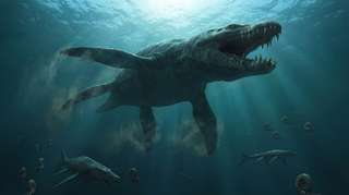
On le croyait roi des océans au Jurassique : une vertèbre percée raconte un combat d’une violence extrême
os dent vertèbre chasse musée Jurassique fossile Ichthyosauria Ichthyosaurus Pliosaurus comportement
Oublié pendant plus d’un siècle dans les tiroirs d’un musée, un fossile refait aujourd’hui surface et raconte une scène de violence extrême dans les océans du Jurassique. Une vertèbre d’ichtyosaure, marquée par l’impact d’une dent brisée, révèle peut-être l’attaque d’un des plus redoutables...
17/06/2026 futura-terre
Description d'un fossile de dinosaure rare du groupe de Nanaimo du Crétacé supérieur
os vertèbre description Canada Colombie Crétacé Crétacé supérieur fossile Dinosauria Ornithomimosauria
Les chercheurs ont décrit une seule vertèbre caudale de dinosaure (coccyx) de l'île Denman (Colombie-Britannique, Canada).  Il a été identifié comme une vertèbre caudale d'ornithomimosaure. Le fossile, censé représenter un os de la partie médiane de la queue, n'est que le deuxième fossile de dinosaure identifié dans le groupe de Nanaimo du Crétacé supérieur. De plus, il
10/06/2026 everythingdinosaur ⚙ Traduction automatique
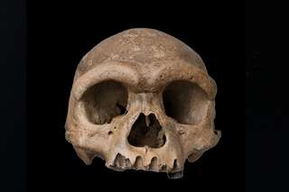
Les scientifiques pensaient les Denisoviens introuvables : cette découverte spectaculaire change désormais tout
os ADN Chine découverte Homo sapiens origine humaine crâne
Pendant quinze ans, les Denisoviens n'ont eu ni visage ni contour. Juste de l'ADN extrait d'un fragment d'os sibérien grand comme un ongle. En 2025, un crâne exceptionnel découvert en Chine a tout changé. Homo longi, surnommé l'Homme dragon, donne enfin un visage à ce groupe humain fantôme. 
06/06/2026 futura-terre
Une grotte vieille d’un million d’années révèle un monde disparu bien avant l’arrivée de l’humain
os Nouvelle-Zélande oiseau
Une grotte près de Waitomo, en Nouvelle-Zélande, vient de livrer l'un des trésors paléontologiques les plus inattendus de ces dernières années. Seize espèces fossilisées, dont un ancêtre inconnu du kākāpō, dormaient là depuis un million d'années. Ce que ces ossements racontent dépasse largement...
04/06/2026 futura-terre
1 2 3 4 5 6 7 8 9 10 11