bone
Anatomical part62 image(s) · 30 News
Image gallery


![Mounted replica of a composite skeleton of Edmontosaurus annectens on display at the University of Oxford Museum, Oxford, England. The original skeleton is compiled from disarticulated fossil bones from a bonebed of the Hell Creek Formation, exposed in the Ruth Mason Quarry in Harding County, South Dakota. It is 8.5 m (28 ft.) long and the skull is almost 1 m (39 in.) in length.[1][2]
↑ Dinosaurs in the Museum. Oxford University Museum of Natural History (brochure, PDF), p. 7
↑ BHI Fossil Replica Catalog 2012. Black Hills Institute of Geological Research, Inc., Hill City, SD, 2012 (PDF), p. 22](https://upload.wikimedia.org/wikipedia/commons/thumb/5/5c/Oxford_Edmontosaurus.jpg/330px-Oxford_Edmontosaurus.jpg)


News
04/06/2026
futura-terre
⚙ Auto-translated

22/05/2026
sciencesetavenir
⚙ Auto-translated

20/05/2026
sci-news
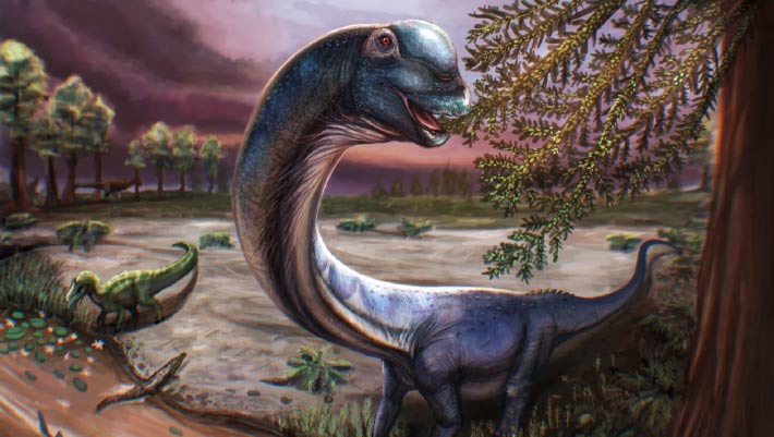
bone
Thailand
Cretaceous
Early Cretaceous
Dinosauria
Somphospondyli
Titanosauriformes
discovery
new species
14/05/2026
sci-news