←

os

Partie anatomique

76 image(s) · 55 Actualités

Galerie d'images

Cuspicephalus scarfi MJMG K1918. A, Original skull on slab of mudstone, Lacking the mandible and dentition; B, outline diagram of preserved bone. Light grey is bone, dark grey is fibrous bone of sagittal crest, black is dental alveoli where unambiguous; C restoration of skull outline, with hypothetical lower jaw. Scale bar = 50 mm. Abbreviation: j, jugal; naof, nasoantorbital fenestra; m, maxilla; o, orbit; pm, premaxilla; q, quadrate; sc,

sagittal crest; sq, squamosal; tr np, trace of nasal process.
Taxons Cuspicephalus

Cuspicephalus scarfi MJMG K1918. A, Original skull on slab of mudstone, Lacking the mandible and dentition; B, outline diagram of preserved bone. Light grey is bone, dark grey is fibrous bone of sagittal crest, black is dental alveoli where unambiguous; C restoration of skull outline, with hypothetical lower jaw. Scale bar = 50 mm. Abbreviation: j, jugal; naof, nasoantorbital fenestra; m, maxilla; o, orbit; pm, premaxilla; q, quadrate; sc, sagittal crest; sq, squamosal; tr np, trace of nasal process.

os crête écaille Cuspicephalus +1
Reconstructed skeleton of a megaraptoran highlighting the known bones of Joaquinraptor casali in blue. Reconstruction by TK Robinson and Andrew McAfee. Scale bar=1 m.
Taxons Joaquinraptor

Reconstructed skeleton of a megaraptoran highlighting the known bones of Joaquinraptor casali in blue. Reconstruction by TK Robinson and Andrew McAfee. Scale bar=1 m.

os écaille Joaquinraptor Megaraptora +1
Photographs of Melkamter pateko (MPEF-PV 11530), visible in medial views on the main block (1) with inset of the tooth (1B) and counterslab (2);. Rendered CT scan detail images (dashed areas represent visible bone that was too thin to be captured by CT scan) of the cranial fragment (1A), manual metacarpal (1C) and dorsal vertebrae (1D–F). Abbreviations: a = alveoli; if = infratemporal fenestra; j = jugal; l = lacrimal; m = maxilla; o = orbit; naof = Nnasoantorbital fenestra; np = nasal process; pm = premaxilla; po = postorbital; q = quadrate; qj = quadratojugal; s = squamosal; t = tooth; vas = vestigial ascending process.
Taxons Melkamter

Photographs of Melkamter pateko (MPEF-PV 11530), visible in medial views on the main block (1) with inset of the tooth (1B) and counterslab (2);. Rendered CT scan detail images (dashed areas represent visible bone that was too thin to be captured by CT scan) of the cranial fragment (1A), manual metacarpal (1C) and dorsal vertebrae (1D–F). Abbreviations: a = alveoli; if = infratemporal fenestra; j = jugal; l = lacrimal; m = maxilla; o = orbit; naof = Nnasoantorbital fenestra; np = nasal process; pm = premaxilla; po = postorbital; q = quadrate; qj = quadratojugal; s = squamosal; t = tooth; vas = vestigial ascending process.

os dent Melkamter tomographie
Tetanuran theropod Cruxicheiros newmanorum gen. et sp. nov. axial vertebrae (WARMS 15771) from the Chipping Norton Limestone Formation,
Bathonian of the United Kingdom. A. Posterior cervical or anterior dorsal vertebra in posterior view. B. Partial middle−posterior dorsal vertebra in right lat−
eral view showing a sagittal cross−section (B1) and in dorsal view (B2). C. Middle−distal caudal vertebra in left lateral (C1) and posterior (C2) views. D. Mid−
dle−posterior dorsal neural arch in anterior (D1), right lateral (D2, D3), and posterior (D4) views. Photographs (A, B1, B2, C1, C2, D1, D2, D4) and line drawing

(D3). Crossed−hatching indicates matrix and grey tone indicates broken bone.
Taxons Cruxicheiros

Tetanuran theropod Cruxicheiros newmanorum gen. et sp. nov. axial vertebrae (WARMS 15771) from the Chipping Norton Limestone Formation, Bathonian of the United Kingdom. A. Posterior cervical or anterior dorsal vertebra in posterior view. B. Partial middle−posterior dorsal vertebra in right lat− eral view showing a sagittal cross−section (B1) and in dorsal view (B2). C. Middle−distal caudal vertebra in left lateral (C1) and posterior (C2) views. D. Mid− dle−posterior dorsal neural arch in anterior (D1), right lateral (D2, D3), and posterior (D4) views. Photographs (A, B1, B2, C1, C2, D1, D2, D4) and line drawing (D3). Crossed−hatching indicates matrix and grey tone indicates broken bone.

os vertèbre dessin Royaume-Uni +4
Skeletal composite of the chilean titanosaur Atacamatitan chilensis. A fragmented sauropod known from the holotype SGO-PV-961, found in the Atacama Desert. The preserved elements consist of:[1]
Right femur, the proximal end of a humerus, two dorsal vertebrae, posterior caudal vertebrae, dorsal ribs and a possibly fragmentary element of the sternum, other fragmented caudal vertebrae and indeterminate bones.[1]
Due to the fragmented nature of the holotype, most cladistic analyzes exclude Atacamatitan. Nevertheless, in 2012, Rubilar-Rogers and Gutstein conducted a preliminary cladistic analysis which placed Atacamatitan within the Lithostrotia.[2]
The original description lacks a lateral view for the humerus and femur, the lateral view for the humerus was based on Alamosaurus and Opisthocoelicaudia, since it shows resemblance with them. The lateral view of the femur is based on Mendozasaurus for the same reason.

Color Key
  Known
  Unknown
Taxons Mendozasaurus

Skeletal composite of the chilean titanosaur Atacamatitan chilensis. A fragmented sauropod known from the holotype SGO-PV-961, found in the Atacama Desert. The preserved elements consist of:[1] Right femur, the proximal end of a humerus, two dorsal vertebrae, posterior caudal vertebrae, dorsal ribs and a possibly fragmentary element of the sternum, other fragmented caudal vertebrae and indeterminate bones.[1] Due to the fragmented nature of the holotype, most cladistic analyzes exclude Atacamatitan. Nevertheless, in 2012, Rubilar-Rogers and Gutstein conducted a preliminary cladistic analysis which placed Atacamatitan within the Lithostrotia.[2] The original description lacks a lateral view for the humerus and femur, the lateral view for the humerus was based on Alamosaurus and Opisthocoelicaudia, since it shows resemblance with them. The lateral view of the femur is based on Mendozasaurus for the same reason. Color Key   Known   Unknown

os humérus description holotype +6
Oneirosaurus caballeroi holotype IGMp879524. A, photograph, drawing and interpretative scheme of the specimen without left mandibular ramus, in right lateral view. B, photograph, drawing and interpretative scheme of the specimen without left mandibular
ramus, in left lateral view. Gray fill, sediment; pattern fill, broken bone surface; black, empty space. Scale bars: A = 50 mm; C-D = 30 mm. Abbreviations: an, angular; avc, anterior aperture of the vidian canal; bo, basioccipital; bptm, basipterygoid meniscus; bs, basisphenoid; c, coronoid; d, dentary; ec, ectopterygoid; ep, epipterygoid; ex, exoccipital; f, frontal; im, internal auditory meatus; is, interorbital septum; j, jugal; la, lacrimal; m, mandible; mx, maxilla; na, nasal; op, opisthotic-exoccipital; p, parietal; par, prearticular; pl, palatine; pmx, premaxilla; pof, postorbitofrontal; pr, prootic; prf, prefrontal; ps, parasphenoid; pt, pterygoid; pvc, posterior aperture of the vidian canal q; quadrate; rv, right vomer; sa, surangular; scp, sclerotic plate; smx, septomaxilla; so, supraoccipital; sp, splenial; stp, stapes; VII, IX-XII, exit of cranial nerves.
Taxons Oneirosaurus

Oneirosaurus caballeroi holotype IGMp879524. A, photograph, drawing and interpretative scheme of the specimen without left mandibular ramus, in right lateral view. B, photograph, drawing and interpretative scheme of the specimen without left mandibular ramus, in left lateral view. Gray fill, sediment; pattern fill, broken bone surface; black, empty space. Scale bars: A = 50 mm; C-D = 30 mm. Abbreviations: an, angular; avc, anterior aperture of the vidian canal; bo, basioccipital; bptm, basipterygoid meniscus; bs, basisphenoid; c, coronoid; d, dentary; ec, ectopterygoid; ep, epipterygoid; ex, exoccipital; f, frontal; im, internal auditory meatus; is, interorbital septum; j, jugal; la, lacrimal; m, mandible; mx, maxilla; na, nasal; op, opisthotic-exoccipital; p, parietal; par, prearticular; pl, palatine; pmx, premaxilla; pof, postorbitofrontal; pr, prootic; prf, prefrontal; ps, parasphenoid; pt, pterygoid; pvc, posterior aperture of the vidian canal q; quadrate; rv, right vomer; sa, surangular; scp, sclerotic plate; smx, septomaxilla; so, supraoccipital; sp, splenial; stp, stapes; VII, IX-XII, exit of cranial nerves.

os écaille dessin holotype +2
Skull in dorsal (a and b, photograph and drawing respectively), and left lateral (c and d, photograph and drawing respectively) views. (e) Premaxillary tooth; (f,g) maxillary teeth (g inverted). amf, anterior maxillary fossa; aof, antorbital fossa; aso, anterior supraorbital; d, dentary; ef, elliptical fossa; f, foramina; fr, frontal; ift, infratemporal fenestra; j, jugal; mx, maxilla; n, nasals; o, orbit; pd, predentary; pdb, postdentary bones; pmx, premaxilla; po, postorbital; pso: posterior supraorbital; prf, prefrontal; qj, quadratojugal; sq, squamosal; stf, supratemporal fenestra. 1–7 denticles. The drawings were processed using Adobe Photoshop CS2 Serial Number: 1045-1412-5685-1654-6343-1431.
Taxons Isaberrysaura

Skull in dorsal (a and b, photograph and drawing respectively), and left lateral (c and d, photograph and drawing respectively) views. (e) Premaxillary tooth; (f,g) maxillary teeth (g inverted). amf, anterior maxillary fossa; aof, antorbital fossa; aso, anterior supraorbital; d, dentary; ef, elliptical fossa; f, foramina; fr, frontal; ift, infratemporal fenestra; j, jugal; mx, maxilla; n, nasals; o, orbit; pd, predentary; pdb, postdentary bones; pmx, premaxilla; po, postorbital; pso: posterior supraorbital; prf, prefrontal; qj, quadratojugal; sq, squamosal; stf, supratemporal fenestra. 1–7 denticles. The drawings were processed using Adobe Photoshop CS2 Serial Number: 1045-1412-5685-1654-6343-1431.

os dent dessin Isaberrysaura +1
Reconstruction of the Bravoceratops skeleton with known bones marked in white. Specimen: TMM 46015-1 (holotype). Scale bar=2 m. Total length along centra=6.97 m.
Taxons Bravoceratops

Reconstruction of the Bravoceratops skeleton with known bones marked in white. Specimen: TMM 46015-1 (holotype). Scale bar=2 m. Total length along centra=6.97 m.

os écaille holotype spécimen +2
Bone mapping of the skeleton of Plesiopharos moelensis gen. et sp. nov. (ML2302) from the Sinemurian (Lower Jurassic) of São Pedro de Moel (Leiria, Portugal). A. Bones distribution map of the three original extracted blocks (1, 2 and 3) containing ML2302. Arrows with question marks represent that the joining relationship between the blocks is unknown. B. Photographs of the five sub-blocks (1A, 1B, 2C, 2D, and 3E) after preparation works.
Taxons Plesiopharos

Bone mapping of the skeleton of Plesiopharos moelensis gen. et sp. nov. (ML2302) from the Sinemurian (Lower Jurassic) of São Pedro de Moel (Leiria, Portugal). A. Bones distribution map of the three original extracted blocks (1, 2 and 3) containing ML2302. Arrows with question marks represent that the joining relationship between the blocks is unknown. B. Photographs of the five sub-blocks (1A, 1B, 2C, 2D, and 3E) after preparation works.

os Portugal Jurassique Sinémurien +2
A color-coded skull diagram of Palatodonta bleekeri, mostly based on a diagram published by Neenan et al. (2013). Some interpretations (such as the presence of a quadratojugal) are based on Maisch (2020). Depicted in left lateral view, mirrored from the original specimen as preserved in right lateral view. Dotted lines indicate missing bone.
Taxons Palatodonta

A color-coded skull diagram of Palatodonta bleekeri, mostly based on a diagram published by Neenan et al. (2013). Some interpretations (such as the presence of a quadratojugal) are based on Maisch (2020). Depicted in left lateral view, mirrored from the original specimen as preserved in right lateral view. Dotted lines indicate missing bone.

os spécimen Palatodonta crâne
Skeletal reconstruction of the Spectrovenator ragei holotype (MZSP-PV 833). Recovered bones in blue.
Taxons Spectrovenator

Skeletal reconstruction of the Spectrovenator ragei holotype (MZSP-PV 833). Recovered bones in blue.

os holotype Spectrovenator
DMNH 2021-05-02, holotype left dentary of Ampelognathus coheni, photos and labeled outline drawings in A, lateral; B, medial; C, dorsal; D, ventral; E, anterior; and F, posterior views. Abbreviations: a#, alveolus and numerical position in tooth row; anf, angular facet; cof, coronoid facet; cop, coronoid process; d6, dentary tooth 6 base; ds, dentary symphysis; mc, Meckelian canal; mg, Meckelian groove; pdd, dorsal facet for predentary; pdv, ventral facet for predentary; saf, surangular facet. Gray fill indicates recessed surfaces. Stippling indicates visible sediment. Cross-hatching indicates broken bone surface. Scale bars equal 5 mm.
Taxons Ampelognathus

DMNH 2021-05-02, holotype left dentary of Ampelognathus coheni, photos and labeled outline drawings in A, lateral; B, medial; C, dorsal; D, ventral; E, anterior; and F, posterior views. Abbreviations: a#, alveolus and numerical position in tooth row; anf, angular facet; cof, coronoid facet; cop, coronoid process; d6, dentary tooth 6 base; ds, dentary symphysis; mc, Meckelian canal; mg, Meckelian groove; pdd, dorsal facet for predentary; pdv, ventral facet for predentary; saf, surangular facet. Gray fill indicates recessed surfaces. Stippling indicates visible sediment. Cross-hatching indicates broken bone surface. Scale bars equal 5 mm.

os écaille dent dessin +2
A skull reconstruction and cranial bones from Shaochilong maortuensis, a small-bodied mid Cretaceous (Turonian) carcharodontosauri-an theropod from Inner Mongolia, China. A, skull reconstruction (courtesy of Brett Booth); B, right maxilla in lateral view (IVPP V.2885.4); C, braincase and skull roof in dorsal view (IVPP V.2885.1-2). Scale bars equal 5 centimeters.
Taxons Shaochilong

A skull reconstruction and cranial bones from Shaochilong maortuensis, a small-bodied mid Cretaceous (Turonian) carcharodontosauri-an theropod from Inner Mongolia, China. A, skull reconstruction (courtesy of Brett Booth); B, right maxilla in lateral view (IVPP V.2885.4); C, braincase and skull roof in dorsal view (IVPP V.2885.1-2). Scale bars equal 5 centimeters.

os écaille Chine Mongolie +4
Abelisaurid theropod Kryptops palaios gen. et sp. nov. MNN GAD1−1 from the Lower Cretaceous Elrhaz Formation of Niger. Left maxilla in lateral

view; stereopair (A) and line drawing (B). Cross−hatching indicates broken bone; dashed lines indicate estimated edges.
Taxons Kryptops

Abelisaurid theropod Kryptops palaios gen. et sp. nov. MNN GAD1−1 from the Lower Cretaceous Elrhaz Formation of Niger. Left maxilla in lateral view; stereopair (A) and line drawing (B). Cross−hatching indicates broken bone; dashed lines indicate estimated edges.

os dessin Niger Crétacé +3
(A) Skeletal reconstruction in left lateral view (missing and damaged portions of the bones in gray). (B) Left ilium in lateral view. (C) Proximal caudal vertebrae in left lateral view with close-up of the infraprezygapophyses. (D) Chevron in cranial view. (E-F) Right scapula in dorsal (E) and lateral (F) views. (G) Last sacral and the two proximalmost caudals in left lateral view. (H) Right pubis in medial view. (I) Right ischium in lateral view. (J) Right femur in distal view. (K) Left metatarsus and distal tarsals in proximal view. (L) Right femur in cranial view. (M-N) Left metatarsus in lateral (M) and dorsal (N) views. Abbreviations: acr, acromion process; ant, antitrochanter; ch, chevron; cv, caudal vertebra(e); diprf, dorsal infraprezygapophyseal fossa; dt, distal tarsal(s); fct, cranial trochanter of femur; fh, femoral head; gl, glenoid fossa; idf, infradiapophyseal fossa; lc, lateral condyle; mc, medial condyle; mep, medial epicondyle; miprf, middle infraprezygapophyseal fossa; mt II, metatarsal II; mt IV, metatarsal IV; mt V, metatarsal V; ns, neural spine; obp, obturator process; pra, preacetabular process; pup, pubic peduncle; sprf, supraprezygapophyseal fossa; sv, sacral vertebra; tfc, tibiofibular crest; tp, transverse process; viprf, ventral infraprezygapophyseal fossa. Scale bars equal 10 cm in (A); 1 cm in (B-N).
https://doi.org/10.1371/journal.pone.0210867.g004
Taxons Gobiraptor

(A) Skeletal reconstruction in left lateral view (missing and damaged portions of the bones in gray). (B) Left ilium in lateral view. (C) Proximal caudal vertebrae in left lateral view with close-up of the infraprezygapophyses. (D) Chevron in cranial view. (E-F) Right scapula in dorsal (E) and lateral (F) views. (G) Last sacral and the two proximalmost caudals in left lateral view. (H) Right pubis in medial view. (I) Right ischium in lateral view. (J) Right femur in distal view. (K) Left metatarsus and distal tarsals in proximal view. (L) Right femur in cranial view. (M-N) Left metatarsus in lateral (M) and dorsal (N) views. Abbreviations: acr, acromion process; ant, antitrochanter; ch, chevron; cv, caudal vertebra(e); diprf, dorsal infraprezygapophyseal fossa; dt, distal tarsal(s); fct, cranial trochanter of femur; fh, femoral head; gl, glenoid fossa; idf, infradiapophyseal fossa; lc, lateral condyle; mc, medial condyle; mep, medial epicondyle; miprf, middle infraprezygapophyseal fossa; mt II, metatarsal II; mt IV, metatarsal IV; mt V, metatarsal V; ns, neural spine; obp, obturator process; pra, preacetabular process; pup, pubic peduncle; sprf, supraprezygapophyseal fossa; sv, sacral vertebra; tfc, tibiofibular crest; tp, transverse process; viprf, ventral infraprezygapophyseal fossa. Scale bars equal 10 cm in (A); 1 cm in (B-N). https://doi.org/10.1371/journal.pone.0210867.g004

os crête écaille vertèbre +1
Skeletal reconstruction of Dineobellator notohesperus gen. et sp. nov., SMP VP-2430, with known elements colored in white. Figured bones are as follows: fused distal caudal vertebra (A); middle caudal vertebra (B); caudal vertebra 1 (C); right femur (D); rib (E); right basipterygoid (F); left lacrimal (reversed) (G); right jugal (H); right humerus (I); right ulna (J); right metacarpal III (K); right manual ungual II (L); right metatarsal II (M); right metatarsal III. (N) Individual scale bars, 2 cm. Skeletal drawing based off work of Scott Hartman.
Taxons Dineobellator

Skeletal reconstruction of Dineobellator notohesperus gen. et sp. nov., SMP VP-2430, with known elements colored in white. Figured bones are as follows: fused distal caudal vertebra (A); middle caudal vertebra (B); caudal vertebra 1 (C); right femur (D); rib (E); right basipterygoid (F); left lacrimal (reversed) (G); right jugal (H); right humerus (I); right ulna (J); right metacarpal III (K); right manual ungual II (L); right metatarsal II (M); right metatarsal III. (N) Individual scale bars, 2 cm. Skeletal drawing based off work of Scott Hartman.

os humérus écaille vertèbre +2
1 2 3 4 5

Actualités

Une côte cassée du plus grand T. rex du monde cache un secret vieux de 66 millions d'années
os métabolisme pathologie fossile Dinosauria Tyrannosaurus squelette
Une côte cassée de Scotty, le plus grand squelette connu de T. rex, contient un extraordinaire réseau fossilisé de vaisseaux sanguins formés pendant la guérison du dinosaure. Les scientifiques ont utilisé l’imagerie neutronique et les rayons X pour examiner l’intérieur de l’os vieux de 66 millions d’années sans l’endommager, révélant des détails rarement conservés dans les fossiles.
24/09/2026 sciencedaily ⚙ Traduction automatique
Des scientifiques découvrent le premier diplodocus jamais découvert en dehors de l'Amérique du Nord
os Espagne Jurassique Jurassique supérieur fossile Dinosauria Diplodocia
Un fossile de dinosaure vieux de 150 millions d'années provenant d'Espagne a été identifié comme le premier Diplodocus confirmé jamais découvert en dehors de l'Amérique du Nord. Le sauropode géant mesurait environ 25 mètres de long et était suffisamment bien conservé pour que les chercheurs puissent associer les caractéristiques distinctives de ses os de la queue au célèbre genre. Son apparition inattendue en Europe suggère que les dinosaures se déplaçaient entre les continents à la fin du Jurassique, peut-être via des connexions terrestres temporaires.
19/09/2026 sciencedaily ⚙ Traduction automatique
Les princesses égyptiennes antiques étaient enterrées avec des armes. Leurs os révèlent pourquoi
os chasse
Les armes enterrées avec les anciennes princesses égyptiennes étaient peut-être plus que des décorations de cérémonie. Les chercheurs étudiant les momies royales de Dahshur ont découvert des changements osseux correspondant à des années de tir à l'arc et d'autres utilisations intenses d'armes. Plusieurs femmes présentaient également des signes de guérison de leurs blessures, ce qui suggère un mode de vie étonnamment actif pouvant inclure la chasse ou l'entraînement militaire.
30/08/2026 sciencedaily-fossils ⚙ Traduction automatique
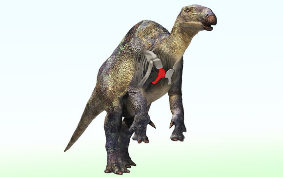
Fossil of Small Duck-Billed Dinosaur Relative Found in Japan
Un fossile d'un petit dinosaure à bec de canard découvert au Japon
os Japon Crétacé fossile Canardia Dinosauria Iguanodontia Styracosterna
Un os fossilisé découvert il y a vingt ans dans le nord-est du Japon appartient à l'un des plus petits dinosaures iguanodontiens jamais découverts dans les roches marines du Crétacé du pays, selon une nouvelle recherche menée par l'Université de Tsukuba. L'article Un fossile d'un petit dinosaure à bec de canard trouvé au Japon est apparu en premier sur Sci.News : Breaking Science News.
20/08/2026 sci-news ⚙ Traduction automatique
Une nouvelle espèce de la période glaciaire se cachait dans les fossiles des fosses de la Brea depuis des décennies
os Pléistocène fossile découverte nouvelle espèce
Les scientifiques ont découvert une nouvelle espèce d’amphibien éteinte cachée dans les collections de fossiles de La Brea Tar Pits à Los Angeles. Nommé Spea labreae, le crapaud de la période glaciaire est une découverte exceptionnellement rare : un seul autre amphibien éteint du Pléistocène a jamais été identifié en Amérique du Nord. Ses ossements étaient conservés dans des collections depuis les années 1950 avant qu'un chercheur ne remarque des caractéristiques qui le distinguaient des espèces connues.
16/08/2026 sciencedaily-paleo ⚙ Traduction automatique
1 2 3 4 5 6 7 8 9 10 11