os
Partie anatomique62 image(s) · 30 Actualités
Galerie d'images


Actualités
04/06/2026
futura-terre

22/05/2026
sciencesetavenir

20/05/2026
sci-news
⚙ Traduction automatique
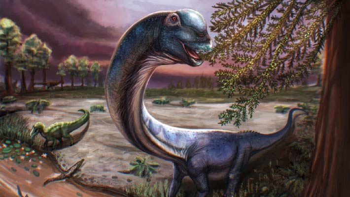
os
Thaïlande
Crétacé
Crétacé inférieur
Dinosauria
Somphospondyli
Titanosauriformes
découverte
nouvelle espèce
14/05/2026
sci-news
⚙ Traduction automatique