os

Partie anatomique

76 image(s) · 52 Actualités

Galerie d'images

Oneirosaurus caballeroi holotype IGMp879524. A, photograph, drawing and interpretative scheme of the specimen without left mandibular ramus, in right lateral view. B, photograph, drawing and interpretative scheme of the specimen without left mandibular
ramus, in left lateral view. Gray fill, sediment; pattern fill, broken bone surface; black, empty space. Scale bars: A = 50 mm; C-D = 30 mm. Abbreviations: an, angular; avc, anterior aperture of the vidian canal; bo, basioccipital; bptm, basipterygoid meniscus; bs, basisphenoid; c, coronoid; d, dentary; ec, ectopterygoid; ep, epipterygoid; ex, exoccipital; f, frontal; im, internal auditory meatus; is, interorbital septum; j, jugal; la, lacrimal; m, mandible; mx, maxilla; na, nasal; op, opisthotic-exoccipital; p, parietal; par, prearticular; pl, palatine; pmx, premaxilla; pof, postorbitofrontal; pr, prootic; prf, prefrontal; ps, parasphenoid; pt, pterygoid; pvc, posterior aperture of the vidian canal q; quadrate; rv, right vomer; sa, surangular; scp, sclerotic plate; smx, septomaxilla; so, supraoccipital; sp, splenial; stp, stapes; VII, IX-XII, exit of cranial nerves.
Taxons Oneirosaurus

Oneirosaurus caballeroi holotype IGMp879524. A, photograph, drawing and interpretative scheme of the specimen without left mandibular ramus, in right lateral view. B, photograph, drawing and interpretative scheme of the specimen without left mandibular ramus, in left lateral view. Gray fill, sediment; pattern fill, broken bone surface; black, empty space. Scale bars: A = 50 mm; C-D = 30 mm. Abbreviations: an, angular; avc, anterior aperture of the vidian canal; bo, basioccipital; bptm, basipterygoid meniscus; bs, basisphenoid; c, coronoid; d, dentary; ec, ectopterygoid; ep, epipterygoid; ex, exoccipital; f, frontal; im, internal auditory meatus; is, interorbital septum; j, jugal; la, lacrimal; m, mandible; mx, maxilla; na, nasal; op, opisthotic-exoccipital; p, parietal; par, prearticular; pl, palatine; pmx, premaxilla; pof, postorbitofrontal; pr, prootic; prf, prefrontal; ps, parasphenoid; pt, pterygoid; pvc, posterior aperture of the vidian canal q; quadrate; rv, right vomer; sa, surangular; scp, sclerotic plate; smx, septomaxilla; so, supraoccipital; sp, splenial; stp, stapes; VII, IX-XII, exit of cranial nerves.

os écaille dessin holotype +2
Mounted replica of a composite skeleton of Edmontosaurus annectens on display at the University of Oxford Museum, Oxford, England. The original skeleton is compiled from disarticulated fossil bones from a bonebed of the Hell Creek Formation, exposed in the Ruth Mason Quarry in Harding County, South Dakota. It is 8.5 m (28 ft.) long and the skull is almost 1 m (39 in.) in length.[1][2]


↑ Dinosaurs in the Museum. Oxford University Museum of Natural History (brochure, PDF), p. 7

↑ BHI Fossil Replica Catalog 2012. Black Hills Institute of Geological Research, Inc., Hill City, SD, 2012 (PDF), p. 22
Taxons Edmontosaurini

Mounted replica of a composite skeleton of Edmontosaurus annectens on display at the University of Oxford Museum, Oxford, England. The original skeleton is compiled from disarticulated fossil bones from a bonebed of the Hell Creek Formation, exposed in the Ruth Mason Quarry in Harding County, South Dakota. It is 8.5 m (28 ft.) long and the skull is almost 1 m (39 in.) in length.[1][2] ↑ Dinosaurs in the Museum. Oxford University Museum of Natural History (brochure, PDF), p. 7 ↑ BHI Fossil Replica Catalog 2012. Black Hills Institute of Geological Research, Inc., Hill City, SD, 2012 (PDF), p. 22

os musée Hell Creek fossile +10
Mounted replica of a composite skeleton of Edmontosaurus annectens on display at the University of Oxford Museum, Oxford, England. The original skeleton is compiled from disarticulated fossil bones from a bonebed of the Hell Creek Formation, exposed in the Ruth Mason Quarry in Harding County, South Dakota. It is 8.5 m (28 ft.) long and the skull is almost 1 m (39 in.) in length.[1][2]


↑ Dinosaurs in the Museum. Oxford University Museum of Natural History (brochure, PDF), p. 7

↑ BHI Fossil Replica Catalog 2012. Black Hills Institute of Geological Research, Inc., Hill City, SD, 2012 (PDF), p. 22
Taxons Kritosaurini

Mounted replica of a composite skeleton of Edmontosaurus annectens on display at the University of Oxford Museum, Oxford, England. The original skeleton is compiled from disarticulated fossil bones from a bonebed of the Hell Creek Formation, exposed in the Ruth Mason Quarry in Harding County, South Dakota. It is 8.5 m (28 ft.) long and the skull is almost 1 m (39 in.) in length.[1][2] ↑ Dinosaurs in the Museum. Oxford University Museum of Natural History (brochure, PDF), p. 7 ↑ BHI Fossil Replica Catalog 2012. Black Hills Institute of Geological Research, Inc., Hill City, SD, 2012 (PDF), p. 22

os musée Hell Creek fossile +10
Mounted replica of a composite skeleton of Edmontosaurus annectens on display at the University of Oxford Museum, Oxford, England. The original skeleton is compiled from disarticulated fossil bones from a bonebed of the Hell Creek Formation, exposed in the Ruth Mason Quarry in Harding County, South Dakota. It is 8.5 m (28 ft.) long and the skull is almost 1 m (39 in.) in length.[1][2]


↑ Dinosaurs in the Museum. Oxford University Museum of Natural History (brochure, PDF), p. 7

↑ BHI Fossil Replica Catalog 2012. Black Hills Institute of Geological Research, Inc., Hill City, SD, 2012 (PDF), p. 22
Taxons Saurolophini

Mounted replica of a composite skeleton of Edmontosaurus annectens on display at the University of Oxford Museum, Oxford, England. The original skeleton is compiled from disarticulated fossil bones from a bonebed of the Hell Creek Formation, exposed in the Ruth Mason Quarry in Harding County, South Dakota. It is 8.5 m (28 ft.) long and the skull is almost 1 m (39 in.) in length.[1][2] ↑ Dinosaurs in the Museum. Oxford University Museum of Natural History (brochure, PDF), p. 7 ↑ BHI Fossil Replica Catalog 2012. Black Hills Institute of Geological Research, Inc., Hill City, SD, 2012 (PDF), p. 22

os musée Hell Creek fossile +10
Mounted replica of a composite skeleton of Edmontosaurus annectens on display at the University of Oxford Museum, Oxford, England. The original skeleton is compiled from disarticulated fossil bones from a bonebed of the Hell Creek Formation, exposed in the Ruth Mason Quarry in Harding County, South Dakota. It is 8.5 m (28 ft.) long and the skull is almost 1 m (39 in.) in length.[1][2]


↑ Dinosaurs in the Museum. Oxford University Museum of Natural History (brochure, PDF), p. 7

↑ BHI Fossil Replica Catalog 2012. Black Hills Institute of Geological Research, Inc., Hill City, SD, 2012 (PDF), p. 22
Taxons Brachylophosaurini

Mounted replica of a composite skeleton of Edmontosaurus annectens on display at the University of Oxford Museum, Oxford, England. The original skeleton is compiled from disarticulated fossil bones from a bonebed of the Hell Creek Formation, exposed in the Ruth Mason Quarry in Harding County, South Dakota. It is 8.5 m (28 ft.) long and the skull is almost 1 m (39 in.) in length.[1][2] ↑ Dinosaurs in the Museum. Oxford University Museum of Natural History (brochure, PDF), p. 7 ↑ BHI Fossil Replica Catalog 2012. Black Hills Institute of Geological Research, Inc., Hill City, SD, 2012 (PDF), p. 22

os musée Hell Creek fossile +10
Fossil bone of Procerosaurus exogyrarum, possible dinosaur from the Czech Republic. National Museum, Prague (2007).
Taxons Ponerosteus

Fossil bone of Procerosaurus exogyrarum, possible dinosaur from the Czech Republic. National Museum, Prague (2007).

os musée Tchéquie fossile +3
Comparison of the holotype (BRSMG Cg3178, A and C right surangular, BAS specimen) and referred specimen (BRSMG Cg2488, B and D left surangular, Lilstock specimen) of Ichthyotitan severnensis gen. et sp. nov. To ease comparison, A and C have been reversed. A-B. Lateral view of both surangulars showing same unique shape; note the upturned, almost 90-degree angle bend and the spatulate-shaped posterior end. C-D. Medial view of both surangulars displaying same morphology posteriorly; anteriorly the Lilstock specimen (D) has been heavily eroded and distorted along its length. Note the position of an elongated foramen on the lateral surface (A-B), identified as part of the fossa surangularis that passes through the bone into the Meckelian canal. See also the damaged (?)angular that is articulated with the surangular and defined by a continuous groove (?suture) as seen in Fig 2H.
Taxons Ichthyotitan

Comparison of the holotype (BRSMG Cg3178, A and C right surangular, BAS specimen) and referred specimen (BRSMG Cg2488, B and D left surangular, Lilstock specimen) of Ichthyotitan severnensis gen. et sp. nov. To ease comparison, A and C have been reversed. A-B. Lateral view of both surangulars showing same unique shape; note the upturned, almost 90-degree angle bend and the spatulate-shaped posterior end. C-D. Medial view of both surangulars displaying same morphology posteriorly; anteriorly the Lilstock specimen (D) has been heavily eroded and distorted along its length. Note the position of an elongated foramen on the lateral surface (A-B), identified as part of the fossa surangularis that passes through the bone into the Meckelian canal. See also the damaged (?)angular that is articulated with the surangular and defined by a continuous groove (?suture) as seen in Fig 2H.

os holotype spécimen Ichthyotitan
Figure description from paper: "Fig. 3 Wimanius odontopalatus Maisch & Matzke, 1998, holotype, GPIT-PV-76272. Photos courtesy of G. Bindellini (Milano). a The complete holotype. b Detail showing a part of the dentition. c Disarticulated bones from the posterior part of the skull
Taxons Wimanius

Figure description from paper: "Fig. 3 Wimanius odontopalatus Maisch & Matzke, 1998, holotype, GPIT-PV-76272. Photos courtesy of G. Bindellini (Milano). a The complete holotype. b Detail showing a part of the dentition. c Disarticulated bones from the posterior part of the skull

os description holotype Wimaniidae +2
Figure description from paper: "Fig. 3 Wimanius odontopalatus Maisch & Matzke, 1998, holotype, GPIT-PV-76272. Photos courtesy of G. Bindellini (Milano). a The complete holotype. b Detail showing a part of the dentition. c Disarticulated bones from the posterior part of the skull
Taxons Wimaniidae

Figure description from paper: "Fig. 3 Wimanius odontopalatus Maisch & Matzke, 1998, holotype, GPIT-PV-76272. Photos courtesy of G. Bindellini (Milano). a The complete holotype. b Detail showing a part of the dentition. c Disarticulated bones from the posterior part of the skull

os description holotype Wimaniidae +2
Partial skull and anterior cervical vertebrae of unidentified pliosaurid plesiosaurian (MPPL 18797) from the uppermost Callovian–middle Oxfordian of Kaberlaba (Asiago, Italy), in left lateral view. Photograph (A) and explanatory drawing (B). White, preserved bone surfaces; dark grey, preserved margins of skull fenestrae; light grey, eroded bone surface; cross hatching, broken bone.
Taxons Anguanax

Partial skull and anterior cervical vertebrae of unidentified pliosaurid plesiosaurian (MPPL 18797) from the uppermost Callovian–middle Oxfordian of Kaberlaba (Asiago, Italy), in left lateral view. Photograph (A) and explanatory drawing (B). White, preserved bone surfaces; dark grey, preserved margins of skull fenestrae; light grey, eroded bone surface; cross hatching, broken bone.

os dessin Italie Callovien +6
Bone mapping of the skeleton of Plesiopharos moelensis gen. et sp. nov. (ML2302) from the Sinemurian (Lower Jurassic) of São Pedro de Moel (Leiria, Portugal). A. Bones distribution map of the three original extracted blocks (1, 2 and 3) containing ML2302. Arrows with question marks represent that the joining relationship between the blocks is unknown. B. Photographs of the five sub-blocks (1A, 1B, 2C, 2D, and 3E) after preparation works.
Taxons Plesiopharos

Bone mapping of the skeleton of Plesiopharos moelensis gen. et sp. nov. (ML2302) from the Sinemurian (Lower Jurassic) of São Pedro de Moel (Leiria, Portugal). A. Bones distribution map of the three original extracted blocks (1, 2 and 3) containing ML2302. Arrows with question marks represent that the joining relationship between the blocks is unknown. B. Photographs of the five sub-blocks (1A, 1B, 2C, 2D, and 3E) after preparation works.

os Portugal Jurassique Sinémurien +2
Outline and overview of the connected slabs TWE 480000454-A, -B, -D, -E, and -M.
TWE 480000454-A shows a single plate-like girdle bone (is). TWE 480000454-B contains
disarticulated vertebrae from the posterior presacral and sacral region (c1-11, na1-3),
fragments of dorsal ribs (dr1, dr2), elements of gastral ribs (unlabelled), the second ischium
(is), the round, plate-like pubis (pu), and three bones, most likely representing sacral- or
caudal ribs (?cr1-3). TWE 480000454-D, -E, and -M mainly contain dorsal ribs (dr3-9) and
elements of gastral ribs as well as two more isolated halves of neural arches (na4, na5), and a

caudal rib (cr1).
Taxons Pararcus

Outline and overview of the connected slabs TWE 480000454-A, -B, -D, -E, and -M. TWE 480000454-A shows a single plate-like girdle bone (is). TWE 480000454-B contains disarticulated vertebrae from the posterior presacral and sacral region (c1-11, na1-3), fragments of dorsal ribs (dr1, dr2), elements of gastral ribs (unlabelled), the second ischium (is), the round, plate-like pubis (pu), and three bones, most likely representing sacral- or caudal ribs (?cr1-3). TWE 480000454-D, -E, and -M mainly contain dorsal ribs (dr3-9) and elements of gastral ribs as well as two more isolated halves of neural arches (na4, na5), and a caudal rib (cr1).

os Pararcus
A color-coded skull diagram of Palatodonta bleekeri, mostly based on a diagram published by Neenan et al. (2013). Some interpretations (such as the presence of a quadratojugal) are based on Maisch (2020). Depicted in left lateral view, mirrored from the original specimen as preserved in right lateral view. Dotted lines indicate missing bone.
Taxons Palatodonta

A color-coded skull diagram of Palatodonta bleekeri, mostly based on a diagram published by Neenan et al. (2013). Some interpretations (such as the presence of a quadratojugal) are based on Maisch (2020). Depicted in left lateral view, mirrored from the original specimen as preserved in right lateral view. Dotted lines indicate missing bone.

os spécimen Palatodonta crâne
Cuspicephalus scarfi MJMG K1918. A, Original skull on slab of mudstone, Lacking the mandible and dentition; B, outline diagram of preserved bone. Light grey is bone, dark grey is fibrous bone of sagittal crest, black is dental alveoli where unambiguous; C restoration of skull outline, with hypothetical lower jaw. Scale bar = 50 mm. Abbreviation: j, jugal; naof, nasoantorbital fenestra; m, maxilla; o, orbit; pm, premaxilla; q, quadrate; sc,

sagittal crest; sq, squamosal; tr np, trace of nasal process.
Taxons Cuspicephalus

Cuspicephalus scarfi MJMG K1918. A, Original skull on slab of mudstone, Lacking the mandible and dentition; B, outline diagram of preserved bone. Light grey is bone, dark grey is fibrous bone of sagittal crest, black is dental alveoli where unambiguous; C restoration of skull outline, with hypothetical lower jaw. Scale bar = 50 mm. Abbreviation: j, jugal; naof, nasoantorbital fenestra; m, maxilla; o, orbit; pm, premaxilla; q, quadrate; sc, sagittal crest; sq, squamosal; tr np, trace of nasal process.

os crête écaille Cuspicephalus +1
Photographs of Melkamter pateko (MPEF-PV 11530), visible in medial views on the main block (1) with inset of the tooth (1B) and counterslab (2);. Rendered CT scan detail images (dashed areas represent visible bone that was too thin to be captured by CT scan) of the cranial fragment (1A), manual metacarpal (1C) and dorsal vertebrae (1D–F). Abbreviations: a = alveoli; if = infratemporal fenestra; j = jugal; l = lacrimal; m = maxilla; o = orbit; naof = Nnasoantorbital fenestra; np = nasal process; pm = premaxilla; po = postorbital; q = quadrate; qj = quadratojugal; s = squamosal; t = tooth; vas = vestigial ascending process.
Taxons Melkamter

Photographs of Melkamter pateko (MPEF-PV 11530), visible in medial views on the main block (1) with inset of the tooth (1B) and counterslab (2);. Rendered CT scan detail images (dashed areas represent visible bone that was too thin to be captured by CT scan) of the cranial fragment (1A), manual metacarpal (1C) and dorsal vertebrae (1D–F). Abbreviations: a = alveoli; if = infratemporal fenestra; j = jugal; l = lacrimal; m = maxilla; o = orbit; naof = Nnasoantorbital fenestra; np = nasal process; pm = premaxilla; po = postorbital; q = quadrate; qj = quadratojugal; s = squamosal; t = tooth; vas = vestigial ascending process.

os dent Melkamter tomographie
Skeletal reconstruction of the Spectrovenator ragei holotype (MZSP-PV 833). Recovered bones in blue.
Taxons Spectrovenator

Skeletal reconstruction of the Spectrovenator ragei holotype (MZSP-PV 833). Recovered bones in blue.

os holotype Spectrovenator
1 2 3 4 5

Actualités

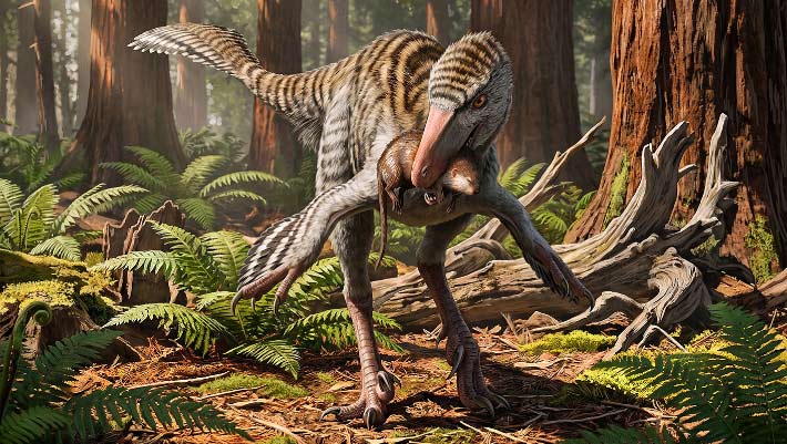
New Troodontid Dinosaur Genus Identified from Fossil Bone Found Decades Ago in New Mexico
Un nouveau genre de dinosaure troodontidé identifié à partir d'un os fossile découvert il y a plusieurs décennies au Nouveau-Mexique
os Mexique fossile Dinevenator Dinosauria Troodontidae oiseau
Les paléontologues ont décrit un nouveau genre de petit dinosaure ressemblant à un oiseau qui vivait dans ce qui est aujourd'hui le Nouveau-Mexique il y a environ 73 millions d'années. L'article Nouveau genre de dinosaures troodontidés identifié à partir d'os fossiles trouvés il y a des décennies au Nouveau-Mexique est apparu en premier sur Sci.News : Breaking Science News.
07/08/2026 sci-news ⚙ Traduction automatique
Le T. rex a-t-il laissé ces marques de morsure vieilles de 66 millions d'années ?
os dent alimentation Dinosauria Tyrannosaurus comportement datation
Les os fossilisés de dinosaures du Wyoming offrent un rare aperçu du comportement alimentaire du Tyrannosaurus rex. Les chercheurs ont examiné plus de 3 000 os datant de 72 à 66 millions d’années et n’en ont trouvé que 12 avec d’éventuelles traces de dents, dont quatre avec des motifs qui correspondaient étroitement à la forme et à l’espacement des dents du T. rex.
05/08/2026 sciencedaily ⚙ Traduction automatique
Célébration de Peter Trusler Palaeoart – Redonner vie aux océans anciens
os fossile Ichthyosauria découverte
L’un des points forts du reportage sur les nouvelles découvertes scientifiques est de voir les œuvres d’art remarquables qui accompagnent de nombreux articles de recherche. Bien que les fossiles en fournissent la preuve, il faut souvent l'habileté d'un paléoartiste talentueux pour transformer les os et les impressions en scènes vivantes des temps profonds. Par exemple, nous avons récemment parlé d’un remarquable fossile d’ichtyosaure.
02/08/2026 everythingdinosaur ⚙ Traduction automatique
Un marteau en os d'éléphant vieux de 500 000 ans montre que les anciens humains étaient des maîtres outilleurs
os découverte
Il y a près de 500 000 ans, les premiers humains d'Angleterre ont transformé un morceau rare d'os d'éléphant en un marteau réutilisable pour aiguiser les outils en pierre. Cette découverte extraordinaire révèle que les anciens outilleurs étaient ingénieux, compétents et capables d’une planification étonnamment avancée.
27/07/2026 sciencedaily ⚙ Traduction automatique
Néandertal semblait manger d’immenses quantités de viande, mais quelque chose ne colle pas
os collagène étude
Des analyses des os de Néandertal révèlent depuis longtemps une énigme alimentaire. Les indices chimiques retrouvés dans leur collagène suggèrent un régime si riche en viande qu'il semble incompatible avec les capacités du corps humain. Une nouvelle étude remet en question une hypothèse...
26/07/2026 futura-terre
1 2 3 4 5 6 7 8 9 10 11