os

Partie anatomique

76 image(s) · 52 Actualités

Galerie d'images

Skeletal reconstruction of Laquintasaura venezuelae, a prionodontian ornithischian (after Barrett et al., 2014). A predentary was restored on the mandible of Laquintasaura, but there is no proof that this bone was present.
Taxons Laquintasaura

Skeletal reconstruction of Laquintasaura venezuelae, a prionodontian ornithischian (after Barrett et al., 2014). A predentary was restored on the mandible of Laquintasaura, but there is no proof that this bone was present.

os Laquintasaura Ornithischia Prionodontia
Holotype of Jakapil kaniukura (MPCA-PV-630), postcranial bones. Speculative silhouette showing preserved elements (a); osteoderm distribution is speculative and partial to show non-osteodermal elements); dorsal vertebra elements in dorsal (b), right lateral (c) and anterior (d,e) views; sacral vertebra in left lateral view (f); mid-caudal vertebra in left lateral view (g); fragment of the mid-shaft of a dorsal rib in posterior view (the enlarged, broken posterior edge is highlighted (h); expanded distal ends of two dorsal ribs (i); left scapula in lateral view (j); right scapula in lateral view (k); right coracoid in lateral view (l); left and right humeri in anterior view (m); probable right ulna in lateral view (n); metacarpals, non-ungual and ungual phalanx in dorsal views (o); left femur elements in anterior view (p); proximal end of the right fibula in lateral view (q); distal end of the left tibia in anterior view (r); ischial elements in side view (s); cervical osteoderms in dorsal view (t), flat scutes in dorsal view (u), spine-like osteoderm in side view (v) and ossicle in dorsal view (w). ac acromial crest, aco asymmetrical cervical osteoderm, alp anterolateral process, ap acromial process, at anterior trochanter, bb basal bone, ebr expanded broken rib edge, di diapophysis, dpc deltopectoral crest, ft fourth trochanter, gl glenoid, mc metacarpals, nc neural canal, ncs neurocentral suture, ph non-ungual phalanx, pp pubic peduncle, poz postzygapophyses, rug marginal rugosities, sb scapular blade, sc scute, tp transverse process, uph ungual phalanx.
Taxons Jakapil

Holotype of Jakapil kaniukura (MPCA-PV-630), postcranial bones. Speculative silhouette showing preserved elements (a); osteoderm distribution is speculative and partial to show non-osteodermal elements); dorsal vertebra elements in dorsal (b), right lateral (c) and anterior (d,e) views; sacral vertebra in left lateral view (f); mid-caudal vertebra in left lateral view (g); fragment of the mid-shaft of a dorsal rib in posterior view (the enlarged, broken posterior edge is highlighted (h); expanded distal ends of two dorsal ribs (i); left scapula in lateral view (j); right scapula in lateral view (k); right coracoid in lateral view (l); left and right humeri in anterior view (m); probable right ulna in lateral view (n); metacarpals, non-ungual and ungual phalanx in dorsal views (o); left femur elements in anterior view (p); proximal end of the right fibula in lateral view (q); distal end of the left tibia in anterior view (r); ischial elements in side view (s); cervical osteoderms in dorsal view (t), flat scutes in dorsal view (u), spine-like osteoderm in side view (v) and ossicle in dorsal view (w). ac acromial crest, aco asymmetrical cervical osteoderm, alp anterolateral process, ap acromial process, at anterior trochanter, bb basal bone, ebr expanded broken rib edge, di diapophysis, dpc deltopectoral crest, ft fourth trochanter, gl glenoid, mc metacarpals, nc neural canal, ncs neurocentral suture, ph non-ungual phalanx, pp pubic peduncle, poz postzygapophyses, rug marginal rugosities, sb scapular blade, sc scute, tp transverse process, uph ungual phalanx.

os vertèbre holotype Jakapil +1
Skull in dorsal (a and b, photograph and drawing respectively), and left lateral (c and d, photograph and drawing respectively) views. (e) Premaxillary tooth; (f,g) maxillary teeth (g inverted). amf, anterior maxillary fossa; aof, antorbital fossa; aso, anterior supraorbital; d, dentary; ef, elliptical fossa; f, foramina; fr, frontal; ift, infratemporal fenestra; j, jugal; mx, maxilla; n, nasals; o, orbit; pd, predentary; pdb, postdentary bones; pmx, premaxilla; po, postorbital; pso: posterior supraorbital; prf, prefrontal; qj, quadratojugal; sq, squamosal; stf, supratemporal fenestra. 1–7 denticles. The drawings were processed using Adobe Photoshop CS2 Serial Number: 1045-1412-5685-1654-6343-1431.
Taxons Isaberrysaura

Skull in dorsal (a and b, photograph and drawing respectively), and left lateral (c and d, photograph and drawing respectively) views. (e) Premaxillary tooth; (f,g) maxillary teeth (g inverted). amf, anterior maxillary fossa; aof, antorbital fossa; aso, anterior supraorbital; d, dentary; ef, elliptical fossa; f, foramina; fr, frontal; ift, infratemporal fenestra; j, jugal; mx, maxilla; n, nasals; o, orbit; pd, predentary; pdb, postdentary bones; pmx, premaxilla; po, postorbital; pso: posterior supraorbital; prf, prefrontal; qj, quadratojugal; sq, squamosal; stf, supratemporal fenestra. 1–7 denticles. The drawings were processed using Adobe Photoshop CS2 Serial Number: 1045-1412-5685-1654-6343-1431.

os dent dessin Isaberrysaura +1
Skeletal reconstruction of Yuxisaurus kopchicki showing some of the main preserved elements from the holotype (highlighted in blue), with details of the skull bones (A), cervical vertebrae (B), dorsal vertebrae (C), left scapula (D), right humerus (E), and left femur (F).
Scale bars equal 5 cm (A–C) or 10 cm (D–F). The facial region and distal scapula are mirrored. Osteoderms have been omitted for convenience.
Taxons Yuxisaurus

Skeletal reconstruction of Yuxisaurus kopchicki showing some of the main preserved elements from the holotype (highlighted in blue), with details of the skull bones (A), cervical vertebrae (B), dorsal vertebrae (C), left scapula (D), right humerus (E), and left femur (F). Scale bars equal 5 cm (A–C) or 10 cm (D–F). The facial region and distal scapula are mirrored. Osteoderms have been omitted for convenience.

os humérus écaille holotype +2
Preserved bones of Sanjuansaurus gordilloi (PVSJ 605), positioned as they were found in the field. Abbreviations: ca1–15, caudal vertebrae 1–15; c2–9, cervical vertebrae 2–9; d1–14, dorsal vertebrae 1–14; l Fe, left femur; l il, left ilium; l MII, left metatarsal II; l Sc, left scapula and coracoid; l Ti, left tibia; l Ul, left ulna; Ph, manual phalanx; R, rib; r As-Ca, right astragalus and calcaneum; r Fe, right femur; r Fi, right fibula; r Fe, right femur; r Mx, right maxilla; r Pu, right pubis; r Sc, right scapula and coracoid; r Ti, right tibia; s1–3, sacral vertebrae 1–3. Scale bars equals 20 cm.
Taxons Sanjuansaurus

Preserved bones of Sanjuansaurus gordilloi (PVSJ 605), positioned as they were found in the field. Abbreviations: ca1–15, caudal vertebrae 1–15; c2–9, cervical vertebrae 2–9; d1–14, dorsal vertebrae 1–14; l Fe, left femur; l il, left ilium; l MII, left metatarsal II; l Sc, left scapula and coracoid; l Ti, left tibia; l Ul, left ulna; Ph, manual phalanx; R, rib; r As-Ca, right astragalus and calcaneum; r Fe, right femur; r Fi, right fibula; r Fe, right femur; r Mx, right maxilla; r Pu, right pubis; r Sc, right scapula and coracoid; r Ti, right tibia; s1–3, sacral vertebrae 1–3. Scale bars equals 20 cm.

os écaille Sanjuansaurus
Representative bones of Pulanesaura eocollum. (a) anterior-to-middle cervical vertebrae (BP/1/6199) in left lateral view; (b) holotypic anterior-most dorsal neural arch (BP/1/6882) in left lateral and anterior views; (c) anterior dorsal neural arch (BP/1/6984) in anterior and right lateral views; (d) anterior mid-dorsal neural arch (BP/1/6183) in anterior and right lateral views; (e) middle dorsal neural arch (BP/1/6770) in posterior view; (f) anterior caudal vertebra (BP/1/6646) in right lateral and posterior views; (g) right humerus (BP/1/6193) in anterior view; (h) left ulna (BP/1/6210) in lateral and proximal views; (i) ?left clavicle (BP/1/6752) in dorsal view; (j) left pedal ungual I (BP/1/6186) in proximal and medial views; (k) left tibia (BP/1/6200) in anterior and lateral views; (l) right ischium (reversed) (BP/1/7366) in lateral view. Abbreviations: aidf, anterior infradiapophyseal fossa; ain, anterior incline of the neural spine; ep, epipophysis; hyp, hyposphene; mr, medial ridge; pp, parapophysis; prdl, prezygodiapophyseal lamina; prz, prezygapophyses; rf, radial fossa; spol, spinopostzygapophyseal lamina; vc, ventral convexity. Scale bars equal 5 cm in a-f and i, j; 10 cm in g, h, k, l. Silhouette drawn by BWM Photographs by BWM.
Taxons Pulanesaura

Representative bones of Pulanesaura eocollum. (a) anterior-to-middle cervical vertebrae (BP/1/6199) in left lateral view; (b) holotypic anterior-most dorsal neural arch (BP/1/6882) in left lateral and anterior views; (c) anterior dorsal neural arch (BP/1/6984) in anterior and right lateral views; (d) anterior mid-dorsal neural arch (BP/1/6183) in anterior and right lateral views; (e) middle dorsal neural arch (BP/1/6770) in posterior view; (f) anterior caudal vertebra (BP/1/6646) in right lateral and posterior views; (g) right humerus (BP/1/6193) in anterior view; (h) left ulna (BP/1/6210) in lateral and proximal views; (i) ?left clavicle (BP/1/6752) in dorsal view; (j) left pedal ungual I (BP/1/6186) in proximal and medial views; (k) left tibia (BP/1/6200) in anterior and lateral views; (l) right ischium (reversed) (BP/1/7366) in lateral view. Abbreviations: aidf, anterior infradiapophyseal fossa; ain, anterior incline of the neural spine; ep, epipophysis; hyp, hyposphene; mr, medial ridge; pp, parapophysis; prdl, prezygodiapophyseal lamina; prz, prezygapophyses; rf, radial fossa; spol, spinopostzygapophyseal lamina; vc, ventral convexity. Scale bars equal 5 cm in a-f and i, j; 10 cm in g, h, k, l. Silhouette drawn by BWM Photographs by BWM.

os humérus vertèbre Pulanesaura
Fig. 5. Forelimb bones of the titanosaurian sauropod Bustingorrytitan shiva gen. et sp. nov. from “Bustingorry II” site, Neuquén Province, Argentina,
upper Cenomanian. A. Left coracoid (holotype, MMCH-Pv 59/13) in lateral view. B. Left scapula (holotype, MMCH-Pv 59/11) in lateral view. C. Left
humerus (holotype, MMCH-Pv 59/21) in proximal (C1), anterior (C2), and distal (C3) views. D. Right radius (holotype, MMCH-Pv 59/22) in posterior
view. E. Left sternal plate (holotype, MMCH-Pv 59/15) in dorsal view. F. Articulated metacarpals I–V (holotype, MMCH-Pv 59/25–29) in proximal
(F1), anterior (F2) and distal (F3) views. G. Right ulna (holotype, MMCH-Pv 59/23) in medial (G1) and proximal (G2) views. Abbreviations: cf, coracoid

foramen; gas, glenoid articular surface; igl, infraglenoid lip; I–V, metacarpals. Scale bars 200 mm.
Taxons Bustingorrytitan

Fig. 5. Forelimb bones of the titanosaurian sauropod Bustingorrytitan shiva gen. et sp. nov. from “Bustingorry II” site, Neuquén Province, Argentina, upper Cenomanian. A. Left coracoid (holotype, MMCH-Pv 59/13) in lateral view. B. Left scapula (holotype, MMCH-Pv 59/11) in lateral view. C. Left humerus (holotype, MMCH-Pv 59/21) in proximal (C1), anterior (C2), and distal (C3) views. D. Right radius (holotype, MMCH-Pv 59/22) in posterior view. E. Left sternal plate (holotype, MMCH-Pv 59/15) in dorsal view. F. Articulated metacarpals I–V (holotype, MMCH-Pv 59/25–29) in proximal (F1), anterior (F2) and distal (F3) views. G. Right ulna (holotype, MMCH-Pv 59/23) in medial (G1) and proximal (G2) views. Abbreviations: cf, coracoid foramen; gas, glenoid articular surface; igl, infraglenoid lip; I–V, metacarpals. Scale bars 200 mm.

os humérus écaille Argentine +4
Bones referred to Ibirania parva, A. Middle caudal vertebra (MPMA 08-0060-07) referred to I. parva. B. Views of partial right ulna (LPP-PV-0202) of the I. parva holotype. According to Navarro et al. (2022).

Bones referred to Ibirania parva, A. Middle caudal vertebra (MPMA 08-0060-07) referred to I. parva. B. Views of partial right ulna (LPP-PV-0202) of the I. parva holotype. According to Navarro et al. (2022).

os vertèbre holotype Ibirania +1
Herbivorous dinosaur found in the Al-khoudh area.  This dinosaur is similar to the Zalmoxes and Rhabdodon dinosaurs.  The skeleton in the Bait Al Baranda Museum was assembled from bones borrowed from several museums.

Herbivorous dinosaur found in the Al-khoudh area. This dinosaur is similar to the Zalmoxes and Rhabdodon dinosaurs. The skeleton in the Bait Al Baranda Museum was assembled from bones borrowed from several museums.

os musée Dinosauria Mochlodon +3
Dinosaur Bones

Dinosaur Bones

os Sri Lanka Anzu Dinosauria
MRF 319, a partial oviraptorosaurian skeleton referred to Anzu wyliei. 
(A) Skeletal reconstruction in left lateral view, with preserved bones in gray and bones represented in other Anzu specimens in white (hatching indicates heavily reconstructed portions of the ilia of CM 78001). Middle-posterior (ninth?) cervical vertebra in (B) anterior, (C) left lateral, and (D) dorsal views. Posterior (11th?) cervical vertebra in (E) anterior, (F) left lateral, and (G) dorsal views. Posterior (12th?) cervical vertebra in (H) anterior, (I) left lateral, and (J) dorsal views. Anteroposteriorly crushed left radius in lateral (K) and anterior (L) views. Mediolaterally crushed left ulna in lateral (M) and anterior (N) views. (O) Partial left scapulocoracoid in lateral view. Dorsal rib in anterior (P) and posterior (Q) views. Abbreviations: acr, acromial process; cr, cervical rib; dip, distal processes; pat, pathology; pf, pneumatic fossa. Scale bars = 50 cm in A; 1 cm in B–Q.

MRF 319, a partial oviraptorosaurian skeleton referred to Anzu wyliei. (A) Skeletal reconstruction in left lateral view, with preserved bones in gray and bones represented in other Anzu specimens in white (hatching indicates heavily reconstructed portions of the ilia of CM 78001). Middle-posterior (ninth?) cervical vertebra in (B) anterior, (C) left lateral, and (D) dorsal views. Posterior (11th?) cervical vertebra in (E) anterior, (F) left lateral, and (G) dorsal views. Posterior (12th?) cervical vertebra in (H) anterior, (I) left lateral, and (J) dorsal views. Anteroposteriorly crushed left radius in lateral (K) and anterior (L) views. Mediolaterally crushed left ulna in lateral (M) and anterior (N) views. (O) Partial left scapulocoracoid in lateral view. Dorsal rib in anterior (P) and posterior (Q) views. Abbreviations: acr, acromial process; cr, cervical rib; dip, distal processes; pat, pathology; pf, pneumatic fossa. Scale bars = 50 cm in A; 1 cm in B–Q.

os écaille vertèbre pathologie +5
MRF 319, a partial oviraptorosaurian skeleton referred to Anzu wyliei. Skeletal reconstruction in left lateral view, with preserved bones in gray and bones represented in other Anzu specimens in white (hatching indicates heavily reconstructed portions of the ilia. Scale bar = 50 cm (19.7 in)

MRF 319, a partial oviraptorosaurian skeleton referred to Anzu wyliei. Skeletal reconstruction in left lateral view, with preserved bones in gray and bones represented in other Anzu specimens in white (hatching indicates heavily reconstructed portions of the ilia. Scale bar = 50 cm (19.7 in)

os écaille spécimen Anzu +3
Osteohistology of UALVP 59606, metatarsal IV of Citipes elegans. Overview of histological thin section of UALVP 59606 in plane-polarized (A) and cross-polarized (B) light, showing locations of close-up images. Detail (C) of cortex showing predominantly primary fibrolamellar bone with longitudinal-reticular vascularity, endosteal layer, and cyclical growth marks (arrows), under plane-polarized light. Close-up (D) of well developed endosteal layer on the medullary cavity, showing sec- ondary excavation by simple vascular canals, under cross-polarized light. Close-up (E) of tightly packed cyclical growth marks (arrows) near the periosteal surface of the cortex, under plane-polarized light. Close-up (F) of periosteal surface of cortex, showing avascular parallel-fibered bone indicating an incipient external fundamental system (double ended arrow) with three LAGs (arrows). Abbreviations: efs, incipient external fundamental system; el, endosteal lamellae; hb, Haversian bone; pfb, parallel-fibered bone; radv, radial simple vascular canal; so, secondary osteon.

Osteohistology of UALVP 59606, metatarsal IV of Citipes elegans. Overview of histological thin section of UALVP 59606 in plane-polarized (A) and cross-polarized (B) light, showing locations of close-up images. Detail (C) of cortex showing predominantly primary fibrolamellar bone with longitudinal-reticular vascularity, endosteal layer, and cyclical growth marks (arrows), under plane-polarized light. Close-up (D) of well developed endosteal layer on the medullary cavity, showing sec- ondary excavation by simple vascular canals, under cross-polarized light. Close-up (E) of tightly packed cyclical growth marks (arrows) near the periosteal surface of the cortex, under plane-polarized light. Close-up (F) of periosteal surface of cortex, showing avascular parallel-fibered bone indicating an incipient external fundamental system (double ended arrow) with three LAGs (arrows). Abbreviations: efs, incipient external fundamental system; el, endosteal lamellae; hb, Haversian bone; pfb, parallel-fibered bone; radv, radial simple vascular canal; so, secondary osteon.

os croissance Citipes Leptorhynchos +1
Main evolutionary steps proposed for the morphofunctional and postural changes of the sauropod pedes. (A) Sauropod body mass through time (in metric tons) based on the sauropod body mass estimations of (41) (NB: data lacking for the second half of the Upper Cretaceous so illustrated here faded, in continuity with the data recorded in the Cretaceous). Schematic outlines of selected large specimens illustrated in the curve, including (from left to right) P. engelhardti, Vulcanodon karibaensis, R. brownei, G. brancai, Cedarosaurus weiskopfae, and Notocolossus gonzalezparejasi. (B) Projected evolutionary changes occurring in the sauropod pes associated with trend in body mass, including 1, skeletal and functional digitigrade pedal posture among basal non-sauropod sauropodomorphs with an incipient soft tissue pad (ISP) (see figs. S34 and S35); 2 and 3, expansion of a well-developed soft tissue pad beneath the elevated pedal bones (SP), resulting in a functionally plantigrade pes + retention of skeletal posture within a range of digitigrady; 4, retention of a soft tissue pad and yet undetermined trend toward more elevated bones; 5, conservation of the neomorphic soft tissue pad within all lineages. Selected examples of well-preserved non-sauropod sauropodomorph and sauropod pedal tracks illustrated above the trends, including (from left to right) Evazoum siriguii; Pseudotetrasauropus bipedoida, Eosauropus isp., Lavinipes cheminii; Kalosauropus pollex, Liujianpus shunan, Polyonyx gomesi; Parabrontopodus mcintoshi; Brontopodus birdi; Titanopodus mendozensis; and unnamed Asian sauropod track. Source of adapted drawing and notes are listed in table S9 and data S2.
Taxons Evazoum

Main evolutionary steps proposed for the morphofunctional and postural changes of the sauropod pedes. (A) Sauropod body mass through time (in metric tons) based on the sauropod body mass estimations of (41) (NB: data lacking for the second half of the Upper Cretaceous so illustrated here faded, in continuity with the data recorded in the Cretaceous). Schematic outlines of selected large specimens illustrated in the curve, including (from left to right) P. engelhardti, Vulcanodon karibaensis, R. brownei, G. brancai, Cedarosaurus weiskopfae, and Notocolossus gonzalezparejasi. (B) Projected evolutionary changes occurring in the sauropod pes associated with trend in body mass, including 1, skeletal and functional digitigrade pedal posture among basal non-sauropod sauropodomorphs with an incipient soft tissue pad (ISP) (see figs. S34 and S35); 2 and 3, expansion of a well-developed soft tissue pad beneath the elevated pedal bones (SP), resulting in a functionally plantigrade pes + retention of skeletal posture within a range of digitigrady; 4, retention of a soft tissue pad and yet undetermined trend toward more elevated bones; 5, conservation of the neomorphic soft tissue pad within all lineages. Selected examples of well-preserved non-sauropod sauropodomorph and sauropod pedal tracks illustrated above the trends, including (from left to right) Evazoum siriguii; Pseudotetrasauropus bipedoida, Eosauropus isp., Lavinipes cheminii; Kalosauropus pollex, Liujianpus shunan, Polyonyx gomesi; Parabrontopodus mcintoshi; Brontopodus birdi; Titanopodus mendozensis; and unnamed Asian sauropod track. Source of adapted drawing and notes are listed in table S9 and data S2.

os tissus Crétacé spécimen +6
Main evolutionary steps proposed for the morphofunctional and postural changes of the sauropod pedes. (A) Sauropod body mass through time (in metric tons) based on the sauropod body mass estimations of (41) (NB: data lacking for the second half of the Upper Cretaceous so illustrated here faded, in continuity with the data recorded in the Cretaceous). Schematic outlines of selected large specimens illustrated in the curve, including (from left to right) P. engelhardti, Vulcanodon karibaensis, R. brownei, G. brancai, Cedarosaurus weiskopfae, and Notocolossus gonzalezparejasi. (B) Projected evolutionary changes occurring in the sauropod pes associated with trend in body mass, including 1, skeletal and functional digitigrade pedal posture among basal non-sauropod sauropodomorphs with an incipient soft tissue pad (ISP) (see figs. S34 and S35); 2 and 3, expansion of a well-developed soft tissue pad beneath the elevated pedal bones (SP), resulting in a functionally plantigrade pes + retention of skeletal posture within a range of digitigrady; 4, retention of a soft tissue pad and yet undetermined trend toward more elevated bones; 5, conservation of the neomorphic soft tissue pad within all lineages. Selected examples of well-preserved non-sauropod sauropodomorph and sauropod pedal tracks illustrated above the trends, including (from left to right) Evazoum siriguii; Pseudotetrasauropus bipedoida, Eosauropus isp., Lavinipes cheminii; Kalosauropus pollex, Liujianpus shunan, Polyonyx gomesi; Parabrontopodus mcintoshi; Brontopodus birdi; Titanopodus mendozensis; and unnamed Asian sauropod track. Source of adapted drawing and notes are listed in table S9 and data S2.
Taxons Kalosauropus

Main evolutionary steps proposed for the morphofunctional and postural changes of the sauropod pedes. (A) Sauropod body mass through time (in metric tons) based on the sauropod body mass estimations of (41) (NB: data lacking for the second half of the Upper Cretaceous so illustrated here faded, in continuity with the data recorded in the Cretaceous). Schematic outlines of selected large specimens illustrated in the curve, including (from left to right) P. engelhardti, Vulcanodon karibaensis, R. brownei, G. brancai, Cedarosaurus weiskopfae, and Notocolossus gonzalezparejasi. (B) Projected evolutionary changes occurring in the sauropod pes associated with trend in body mass, including 1, skeletal and functional digitigrade pedal posture among basal non-sauropod sauropodomorphs with an incipient soft tissue pad (ISP) (see figs. S34 and S35); 2 and 3, expansion of a well-developed soft tissue pad beneath the elevated pedal bones (SP), resulting in a functionally plantigrade pes + retention of skeletal posture within a range of digitigrady; 4, retention of a soft tissue pad and yet undetermined trend toward more elevated bones; 5, conservation of the neomorphic soft tissue pad within all lineages. Selected examples of well-preserved non-sauropod sauropodomorph and sauropod pedal tracks illustrated above the trends, including (from left to right) Evazoum siriguii; Pseudotetrasauropus bipedoida, Eosauropus isp., Lavinipes cheminii; Kalosauropus pollex, Liujianpus shunan, Polyonyx gomesi; Parabrontopodus mcintoshi; Brontopodus birdi; Titanopodus mendozensis; and unnamed Asian sauropod track. Source of adapted drawing and notes are listed in table S9 and data S2.

os tissus Crétacé spécimen +6
Remake of the original picture of the Drzewica Formation. Terrestrial environment of the Pliensbachian-Toarcian boundary of Fennoscandinavia  Inland environment of the  Bornholm Coast, nearby the German realm of the Ciechocinek Formation. Includes
Ciechocinek Formation (Lower Toarcian, Bones) and Drzwica Formation (Latest Pliensbachian, Footprints) Fauna
Sorthat Formation environment, fluvial influenced mainland with Cheirolepidaceae and Bennetitales as dominant flora
Dinosaurs are based on material found on various locations of Northern Germany, and Footprints of the underliying Drzewica Formation at the Holy Cross Mountains, connected with Bornholm at the time.
Dinosaur Species appeared: 

Megalosauripus isp. Large Footprints (+65 cm) found on the Drzewica Formation. There is a dorsal vertebrae on the German Margin of the Ciechocinek Formation assigend to Megalosauria (Huene, 1966).
Gravisauria spp. representing the Grimmen Sauropod reported on 2014, as a taxon related with Tazoudasaurus. Barapasaurus-like footprints are know from the Drzewica Formation.
Coelophysoidea spp. based on coeval Anchisauripus tracks from the Holy Cross Mountains.
Basal Ornithischan, related to Eocursor, based on a crouching trace (Gerard Dariusz Gierlinski, Martin G. Lockley, Grzegorz Niedźwiedzki:2009).
Massospondylidae spp. based on Otozum-like tracks.

Remake of the original picture of the Drzewica Formation. Terrestrial environment of the Pliensbachian-Toarcian boundary of Fennoscandinavia Inland environment of the Bornholm Coast, nearby the German realm of the Ciechocinek Formation. Includes Ciechocinek Formation (Lower Toarcian, Bones) and Drzwica Formation (Latest Pliensbachian, Footprints) Fauna Sorthat Formation environment, fluvial influenced mainland with Cheirolepidaceae and Bennetitales as dominant flora Dinosaurs are based on material found on various locations of Northern Germany, and Footprints of the underliying Drzewica Formation at the Holy Cross Mountains, connected with Bornholm at the time. Dinosaur Species appeared: Megalosauripus isp. Large Footprints (+65 cm) found on the Drzewica Formation. There is a dorsal vertebrae on the German Margin of the Ciechocinek Formation assigend to Megalosauria (Huene, 1966). Gravisauria spp. representing the Grimmen Sauropod reported on 2014, as a taxon related with Tazoudasaurus. Barapasaurus-like footprints are know from the Drzewica Formation. Coelophysoidea spp. based on coeval Anchisauripus tracks from the Holy Cross Mountains. Basal Ornithischan, related to Eocursor, based on a crouching trace (Gerard Dariusz Gierlinski, Martin G. Lockley, Grzegorz Niedźwiedzki:2009). Massospondylidae spp. based on Otozum-like tracks.

os Allemagne Pliensbachien Toarcien +7
1 2 3 4 5

Actualités

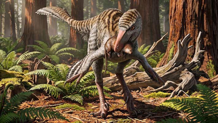
New Troodontid Dinosaur Genus Identified from Fossil Bone Found Decades Ago in New Mexico
Un nouveau genre de dinosaure troodontidé identifié à partir d'un os fossile découvert il y a plusieurs décennies au Nouveau-Mexique
os Mexique fossile Dinevenator Dinosauria Troodontidae oiseau
Les paléontologues ont décrit un nouveau genre de petit dinosaure ressemblant à un oiseau qui vivait dans ce qui est aujourd'hui le Nouveau-Mexique il y a environ 73 millions d'années. L'article Nouveau genre de dinosaures troodontidés identifié à partir d'os fossiles trouvés il y a des décennies au Nouveau-Mexique est apparu en premier sur Sci.News : Breaking Science News.
07/08/2026 sci-news ⚙ Traduction automatique
Le T. rex a-t-il laissé ces marques de morsure vieilles de 66 millions d'années ?
os dent alimentation Dinosauria Tyrannosaurus comportement datation
Les os fossilisés de dinosaures du Wyoming offrent un rare aperçu du comportement alimentaire du Tyrannosaurus rex. Les chercheurs ont examiné plus de 3 000 os datant de 72 à 66 millions d’années et n’en ont trouvé que 12 avec d’éventuelles traces de dents, dont quatre avec des motifs qui correspondaient étroitement à la forme et à l’espacement des dents du T. rex.
05/08/2026 sciencedaily ⚙ Traduction automatique
Célébration de Peter Trusler Palaeoart – Redonner vie aux océans anciens
os fossile Ichthyosauria découverte
L’un des points forts du reportage sur les nouvelles découvertes scientifiques est de voir les œuvres d’art remarquables qui accompagnent de nombreux articles de recherche. Bien que les fossiles en fournissent la preuve, il faut souvent l'habileté d'un paléoartiste talentueux pour transformer les os et les impressions en scènes vivantes des temps profonds. Par exemple, nous avons récemment parlé d’un remarquable fossile d’ichtyosaure.
02/08/2026 everythingdinosaur ⚙ Traduction automatique
Un marteau en os d'éléphant vieux de 500 000 ans montre que les anciens humains étaient des maîtres outilleurs
os découverte
Il y a près de 500 000 ans, les premiers humains d'Angleterre ont transformé un morceau rare d'os d'éléphant en un marteau réutilisable pour aiguiser les outils en pierre. Cette découverte extraordinaire révèle que les anciens outilleurs étaient ingénieux, compétents et capables d’une planification étonnamment avancée.
27/07/2026 sciencedaily ⚙ Traduction automatique
Néandertal semblait manger d’immenses quantités de viande, mais quelque chose ne colle pas
os collagène étude
Des analyses des os de Néandertal révèlent depuis longtemps une énigme alimentaire. Les indices chimiques retrouvés dans leur collagène suggèrent un régime si riche en viande qu'il semble incompatible avec les capacités du corps humain. Une nouvelle étude remet en question une hypothèse...
26/07/2026 futura-terre
1 2 3 4 5 6 7 8 9 10 11