os

Partie anatomique

76 image(s) · 52 Actualités

Galerie d'images

Abelisaurid theropod Kryptops palaios gen. et sp. nov. MNN GAD1−1 from the Lower Cretaceous Elrhaz Formation of Niger. Left maxilla in lateral

view; stereopair (A) and line drawing (B). Cross−hatching indicates broken bone; dashed lines indicate estimated edges.
Taxons Kryptops

Abelisaurid theropod Kryptops palaios gen. et sp. nov. MNN GAD1−1 from the Lower Cretaceous Elrhaz Formation of Niger. Left maxilla in lateral view; stereopair (A) and line drawing (B). Cross−hatching indicates broken bone; dashed lines indicate estimated edges.

os dessin Niger Crétacé +3
Reconstruction of the Abelisaurus skull with original bones of the holotype. Museo Provincial Carlos Ameghino, Cipolletti, Argentina. Scale = 10 cm
Taxons Abelisaurus

Reconstruction of the Abelisaurus skull with original bones of the holotype. Museo Provincial Carlos Ameghino, Cipolletti, Argentina. Scale = 10 cm

os écaille Argentine holotype +3
Reconstructed skull of Duriavenator hesperis showing known material (white) of the holotype and only known specimen. Unknown bones based on related Eustreptospondylus oxoniensis. Scale bar is 10cm, image is 10px/cm. Cranial anatomy based on Benson (2008) "A redescription of "Megalosaurus" hesperis (Dinosauria, Theropoda) from the Inferior Oolite (Bajocian, Middle Jurassic) of Dorset, United Kingdom"
Taxons Duriavenator

Reconstructed skull of Duriavenator hesperis showing known material (white) of the holotype and only known specimen. Unknown bones based on related Eustreptospondylus oxoniensis. Scale bar is 10cm, image is 10px/cm. Cranial anatomy based on Benson (2008) "A redescription of "Megalosaurus" hesperis (Dinosauria, Theropoda) from the Inferior Oolite (Bajocian, Middle Jurassic) of Dorset, United Kingdom"

os écaille Royaume-Uni Bajocien +11
Reconstructed skull of the holotype and only specimen of Leshansaurus qianweiensis based on the possibly related megalosaurid Dubreuillosaurus valesdunensis. Scale bar is 10cm, image is 10px/cm. Cranial anatomy from figures in Fei et al. (2009) "A new carnosaur from the Late Jurassic of Qianwei, Sichuan, China". White bones are figured, light grey bones are unfigured and minimally described.
Taxons Leshansaurus

Reconstructed skull of the holotype and only specimen of Leshansaurus qianweiensis based on the possibly related megalosaurid Dubreuillosaurus valesdunensis. Scale bar is 10cm, image is 10px/cm. Cranial anatomy from figures in Fei et al. (2009) "A new carnosaur from the Late Jurassic of Qianwei, Sichuan, China". White bones are figured, light grey bones are unfigured and minimally described.

os écaille Chine Jurassique +9
Autapomorphies proposed for Diuqin lechiguanae gen. et sp. nov. (MUCPv 1401), with images generated from three-dimensional scans of selected bones. A–B, posteriormost (last) sacral neural arch (MUCPv 1401/1) in dorsal (A) and posterior (B) views. C, anterior caudal neural arch (MUCPv 1401/2) in dorsal view. D, left humerus (MUCPv 1401/4) in lateral view. Abbreviations: al, accessory lamina; dldpr, distolateral deltopectoral ridge; for, foramen. Scale bars equal 3 centimeters
Taxons Diuqin

Autapomorphies proposed for Diuqin lechiguanae gen. et sp. nov. (MUCPv 1401), with images generated from three-dimensional scans of selected bones. A–B, posteriormost (last) sacral neural arch (MUCPv 1401/1) in dorsal (A) and posterior (B) views. C, anterior caudal neural arch (MUCPv 1401/2) in dorsal view. D, left humerus (MUCPv 1401/4) in lateral view. Abbreviations: al, accessory lamina; dldpr, distolateral deltopectoral ridge; for, foramen. Scale bars equal 3 centimeters

os humérus écaille Diuqin +1
Skeletal reconstruction of Dineobellator notohesperus gen. et sp. nov., SMP VP-2430, with known elements colored in white. Figured bones are as follows: fused distal caudal vertebra (A); middle caudal vertebra (B); caudal vertebra 1 (C); right femur (D); rib (E); right basipterygoid (F); left lacrimal (reversed) (G); right jugal (H); right humerus (I); right ulna (J); right metacarpal III (K); right manual ungual II (L); right metatarsal II (M); right metatarsal III. (N) Individual scale bars, 2 cm. Skeletal drawing based off work of Scott Hartman.
Taxons Dineobellator

Skeletal reconstruction of Dineobellator notohesperus gen. et sp. nov., SMP VP-2430, with known elements colored in white. Figured bones are as follows: fused distal caudal vertebra (A); middle caudal vertebra (B); caudal vertebra 1 (C); right femur (D); rib (E); right basipterygoid (F); left lacrimal (reversed) (G); right jugal (H); right humerus (I); right ulna (J); right metacarpal III (K); right manual ungual II (L); right metatarsal II (M); right metatarsal III. (N) Individual scale bars, 2 cm. Skeletal drawing based off work of Scott Hartman.

os humérus écaille vertèbre +2
A diagram showing the bones and feathering preserved in IVPP V 12638, the holotype of Yixianosaurus longimanus, a feathered dinosaur.
Taxons Yixianosaurus

A diagram showing the bones and feathering preserved in IVPP V 12638, the holotype of Yixianosaurus longimanus, a feathered dinosaur.

os holotype Dinosauria Yixianosaurus
(A) Skeletal reconstruction in left lateral view (missing and damaged portions of the bones in gray). (B) Left ilium in lateral view. (C) Proximal caudal vertebrae in left lateral view with close-up of the infraprezygapophyses. (D) Chevron in cranial view. (E-F) Right scapula in dorsal (E) and lateral (F) views. (G) Last sacral and the two proximalmost caudals in left lateral view. (H) Right pubis in medial view. (I) Right ischium in lateral view. (J) Right femur in distal view. (K) Left metatarsus and distal tarsals in proximal view. (L) Right femur in cranial view. (M-N) Left metatarsus in lateral (M) and dorsal (N) views. Abbreviations: acr, acromion process; ant, antitrochanter; ch, chevron; cv, caudal vertebra(e); diprf, dorsal infraprezygapophyseal fossa; dt, distal tarsal(s); fct, cranial trochanter of femur; fh, femoral head; gl, glenoid fossa; idf, infradiapophyseal fossa; lc, lateral condyle; mc, medial condyle; mep, medial epicondyle; miprf, middle infraprezygapophyseal fossa; mt II, metatarsal II; mt IV, metatarsal IV; mt V, metatarsal V; ns, neural spine; obp, obturator process; pra, preacetabular process; pup, pubic peduncle; sprf, supraprezygapophyseal fossa; sv, sacral vertebra; tfc, tibiofibular crest; tp, transverse process; viprf, ventral infraprezygapophyseal fossa. Scale bars equal 10 cm in (A); 1 cm in (B-N).
https://doi.org/10.1371/journal.pone.0210867.g004
Taxons Gobiraptor

(A) Skeletal reconstruction in left lateral view (missing and damaged portions of the bones in gray). (B) Left ilium in lateral view. (C) Proximal caudal vertebrae in left lateral view with close-up of the infraprezygapophyses. (D) Chevron in cranial view. (E-F) Right scapula in dorsal (E) and lateral (F) views. (G) Last sacral and the two proximalmost caudals in left lateral view. (H) Right pubis in medial view. (I) Right ischium in lateral view. (J) Right femur in distal view. (K) Left metatarsus and distal tarsals in proximal view. (L) Right femur in cranial view. (M-N) Left metatarsus in lateral (M) and dorsal (N) views. Abbreviations: acr, acromion process; ant, antitrochanter; ch, chevron; cv, caudal vertebra(e); diprf, dorsal infraprezygapophyseal fossa; dt, distal tarsal(s); fct, cranial trochanter of femur; fh, femoral head; gl, glenoid fossa; idf, infradiapophyseal fossa; lc, lateral condyle; mc, medial condyle; mep, medial epicondyle; miprf, middle infraprezygapophyseal fossa; mt II, metatarsal II; mt IV, metatarsal IV; mt V, metatarsal V; ns, neural spine; obp, obturator process; pra, preacetabular process; pup, pubic peduncle; sprf, supraprezygapophyseal fossa; sv, sacral vertebra; tfc, tibiofibular crest; tp, transverse process; viprf, ventral infraprezygapophyseal fossa. Scale bars equal 10 cm in (A); 1 cm in (B-N). https://doi.org/10.1371/journal.pone.0210867.g004

os vertèbre Gobiraptor
Vertebrae, scapula, forelimb bones, and pelvic bones of Martharaptor greenriverensis (UMNH VP 21400). (A)–Partial cervical neural arch, dorsal view. (B–E)–Cranial dorsal centrum in cranial (B), caudal (C), right lateral (D), and left lateral (E) views. (F–G)–Distal caudal centrum in lateral (F) and ventral (G) views. (H)–Possible ulna. (I)–Possible radius. (J–K)–Left scapula in lateral (J) and medial (K) views. (L)–Proximal end of ischium. (M)–Possible distal end of pubis Scale bar = 50 mm. acr  =  acromium process, ar  =  acetabular rim, gl  =  glenoid, hyp  =  hypapophysis, poz  =  postzygapophysis, pnp  =  pneumatopore, prz  =  prezygapophysis.
Taxons Martharaptor

Vertebrae, scapula, forelimb bones, and pelvic bones of Martharaptor greenriverensis (UMNH VP 21400). (A)–Partial cervical neural arch, dorsal view. (B–E)–Cranial dorsal centrum in cranial (B), caudal (C), right lateral (D), and left lateral (E) views. (F–G)–Distal caudal centrum in lateral (F) and ventral (G) views. (H)–Possible ulna. (I)–Possible radius. (J–K)–Left scapula in lateral (J) and medial (K) views. (L)–Proximal end of ischium. (M)–Possible distal end of pubis Scale bar = 50 mm. acr  =  acromium process, ar  =  acetabular rim, gl  =  glenoid, hyp  =  hypapophysis, poz  =  postzygapophysis, pnp  =  pneumatopore, prz  =  prezygapophysis.

os écaille Martharaptor partiel
Skeletal diagram of Zuolong salleei, based on related basal coelurosaurs. Scale bar = 1 meter. Light colours indicate known material (white is known external bones, light grey is either palate, braincase, or sacrum). Dark colours indicate unknown material (partial bones or unknown braincase)
Taxons Zuolong

Skeletal diagram of Zuolong salleei, based on related basal coelurosaurs. Scale bar = 1 meter. Light colours indicate known material (white is known external bones, light grey is either palate, braincase, or sacrum). Dark colours indicate unknown material (partial bones or unknown braincase)

os écaille Coelurosauria Zuolong +1
(A), silhouette of Maip macrothorax showing the preserved bones in white. (B), reconstruction of the thoracic cavity of Maip at level of D6. (C), interpretative drawing of the excavation of Maip showing the original disposition of the bones. Abbreviations: a, axis; c, coracoid; ind, indeterminate bone; g, gastralia; r, rib; v, vertebrae.
Taxons Maip

(A), silhouette of Maip macrothorax showing the preserved bones in white. (B), reconstruction of the thoracic cavity of Maip at level of D6. (C), interpretative drawing of the excavation of Maip showing the original disposition of the bones. Abbreviations: a, axis; c, coracoid; ind, indeterminate bone; g, gastralia; r, rib; v, vertebrae.

os dessin Maip fouille
Tetanuran theropod Cruxicheiros newmanorum gen. et sp. nov. axial vertebrae (WARMS 15771) from the Chipping Norton Limestone Formation,
Bathonian of the United Kingdom. A. Posterior cervical or anterior dorsal vertebra in posterior view. B. Partial middle−posterior dorsal vertebra in right lat−
eral view showing a sagittal cross−section (B1) and in dorsal view (B2). C. Middle−distal caudal vertebra in left lateral (C1) and posterior (C2) views. D. Mid−
dle−posterior dorsal neural arch in anterior (D1), right lateral (D2, D3), and posterior (D4) views. Photographs (A, B1, B2, C1, C2, D1, D2, D4) and line drawing

(D3). Crossed−hatching indicates matrix and grey tone indicates broken bone.
Taxons Cruxicheiros

Tetanuran theropod Cruxicheiros newmanorum gen. et sp. nov. axial vertebrae (WARMS 15771) from the Chipping Norton Limestone Formation, Bathonian of the United Kingdom. A. Posterior cervical or anterior dorsal vertebra in posterior view. B. Partial middle−posterior dorsal vertebra in right lat− eral view showing a sagittal cross−section (B1) and in dorsal view (B2). C. Middle−distal caudal vertebra in left lateral (C1) and posterior (C2) views. D. Mid− dle−posterior dorsal neural arch in anterior (D1), right lateral (D2, D3), and posterior (D4) views. Photographs (A, B1, B2, C1, C2, D1, D2, D4) and line drawing (D3). Crossed−hatching indicates matrix and grey tone indicates broken bone.

os vertèbre dessin Royaume-Uni +4
Skeleton outline of Dracoraptor hanigani.

Bones highlighted green for present, orange for external moulds and blue for tentatively identified bones. Many unidentified or uncertain elements have been omitted.
Taxons Dracoraptor

Skeleton outline of Dracoraptor hanigani. Bones highlighted green for present, orange for external moulds and blue for tentatively identified bones. Many unidentified or uncertain elements have been omitted.

os Dracoraptor squelette
Reconstruction of the Bravoceratops skeleton with known bones marked in white. Specimen: TMM 46015-1 (holotype). Scale bar=2 m. Total length along centra=6.97 m.
Taxons Bravoceratops

Reconstruction of the Bravoceratops skeleton with known bones marked in white. Specimen: TMM 46015-1 (holotype). Scale bar=2 m. Total length along centra=6.97 m.

os écaille holotype spécimen +2
Ceratopsipes goldenensis Lockley & Hunt, 1995 - ceratopsian dinosaur trackway in the Cretaceous of Colorado, USA.
Ceratopsians are the "horned dinosaurs".  They were large, quadrupedal, herbivorous dinosaurs having a beaked skull and a frill - an extension of bone behind the skull that partially covered the neck.  Ceratopsian dinosaurs are known from the Jurassic and Cretaceous.  The last members of the group died out at the Cretaceous-Tertiary boundary, 65 million years ago.
Seen here is a ceratopsian dinosaur trackway.  Ceratopsian footprints are very rare.  The best examples are in Colorado's Laramie Formation, a nonmarine, coastal plain to deltaic succession of mostly mixed siliciclastic sedimentary rocks - sandstones, siltstones, claystones, and coals.  The footprints here consist of convex bulges on the basal surfaces of structurally-tilted sandstone beds.
The ichnospecies Ceratopsipes goldenensis was named based on footprints at this locality.  The trackmaker was very likely Triceratops, the # 1 most famous ceratopsian dinosaur.


From on-site signage:
Triceratops Tracks
Several tracks of Triceratops, or a closely related horned dinosaur were first discovered in this area and named Ceratopsipes goldenensis (meaning tracks of a horned dinosaur from Golden).  By happy coincidence, the first Triceratops known to science also comes from the greater Denver area near 13th Avenue and Federal Blvd.  Growing to a length of almost 25 feet, Triceratops and other members of the horned dinosaur family were herbivorous animals that roamed in herds.  Their trademark horns and frills that covered their necks probably served for defense.  A replica Triceratops skull is on display in the clubhouse at the Fossil Trace Golf Club.
These tracks (identified by small signs) help us understand that Triceratops walked with a narrow gait and erect front limbs  than with a wide, sprawling gait as previously depicted.  In 1887, the first Triceratops bones known to science were also discovered locally at a site near 13th Avenue and Federal Boulevard in Denver.
Nearby one may see many other traces of life such as burrows, probably made by invertebrates and impressions of plant debris.  Note that you are looking at all tracks and traces from the underside - in negative aspect - what one might call a worm’s eye view.  All of these trace fossils indicate that the Golden area once had a warm, temperate to subtropical climate.


Classification: Animalia, Chordata, Vertebrata, Reptilia, Archosauria, Dinosauria, Ornithischia, Marginocephalia, Ceratopsia, Ceratopsidae
Stratigraphy: Laramie Formation, Maastrichtian Stage, upper Upper Cretaceous
Locality: outcrop along Triceratops Trail, Parfet Prehistoric Preserve, southern side of the town of Golden, Colorado, USA (~vicinity of 39° 44' 35.24" North latitude, 105° 13’ 09.69" West longitude)


Some info. from:

Lockley & Hunt (1995) - Ceratopsid tracks and associated ichnofauna from the Laramie Formation (Upper Cretaceous: Maastrichtian) of Colorado.  Journal of Vertebrate Paleontology 15: 592-614.
Taxons Ceratopsipes

Ceratopsipes goldenensis Lockley & Hunt, 1995 - ceratopsian dinosaur trackway in the Cretaceous of Colorado, USA. Ceratopsians are the "horned dinosaurs". They were large, quadrupedal, herbivorous dinosaurs having a beaked skull and a frill - an extension of bone behind the skull that partially covered the neck. Ceratopsian dinosaurs are known from the Jurassic and Cretaceous. The last members of the group died out at the Cretaceous-Tertiary boundary, 65 million years ago. Seen here is a ceratopsian dinosaur trackway. Ceratopsian footprints are very rare. The best examples are in Colorado's Laramie Formation, a nonmarine, coastal plain to deltaic succession of mostly mixed siliciclastic sedimentary rocks - sandstones, siltstones, claystones, and coals. The footprints here consist of convex bulges on the basal surfaces of structurally-tilted sandstone beds. The ichnospecies Ceratopsipes goldenensis was named based on footprints at this locality. The trackmaker was very likely Triceratops, the # 1 most famous ceratopsian dinosaur. From on-site signage: Triceratops Tracks Several tracks of Triceratops, or a closely related horned dinosaur were first discovered in this area and named Ceratopsipes goldenensis (meaning tracks of a horned dinosaur from Golden). By happy coincidence, the first Triceratops known to science also comes from the greater Denver area near 13th Avenue and Federal Blvd. Growing to a length of almost 25 feet, Triceratops and other members of the horned dinosaur family were herbivorous animals that roamed in herds. Their trademark horns and frills that covered their necks probably served for defense. A replica Triceratops skull is on display in the clubhouse at the Fossil Trace Golf Club. These tracks (identified by small signs) help us understand that Triceratops walked with a narrow gait and erect front limbs than with a wide, sprawling gait as previously depicted. In 1887, the first Triceratops bones known to science were also discovered locally at a site near 13th Avenue and Federal Boulevard in Denver. Nearby one may see many other traces of life such as burrows, probably made by invertebrates and impressions of plant debris. Note that you are looking at all tracks and traces from the underside - in negative aspect - what one might call a worm’s eye view. All of these trace fossils indicate that the Golden area once had a warm, temperate to subtropical climate. Classification: Animalia, Chordata, Vertebrata, Reptilia, Archosauria, Dinosauria, Ornithischia, Marginocephalia, Ceratopsia, Ceratopsidae Stratigraphy: Laramie Formation, Maastrichtian Stage, upper Upper Cretaceous Locality: outcrop along Triceratops Trail, Parfet Prehistoric Preserve, southern side of the town of Golden, Colorado, USA (~vicinity of 39° 44' 35.24" North latitude, 105° 13’ 09.69" West longitude) Some info. from: Lockley & Hunt (1995) - Ceratopsid tracks and associated ichnofauna from the Laramie Formation (Upper Cretaceous: Maastrichtian) of Colorado. Journal of Vertebrate Paleontology 15: 592-614.

os États-Unis Crétacé Jurassique +6
DMNH 2021-05-02, holotype left dentary of Ampelognathus coheni, photos and labeled outline drawings in A, lateral; B, medial; C, dorsal; D, ventral; E, anterior; and F, posterior views. Abbreviations: a#, alveolus and numerical position in tooth row; anf, angular facet; cof, coronoid facet; cop, coronoid process; d6, dentary tooth 6 base; ds, dentary symphysis; mc, Meckelian canal; mg, Meckelian groove; pdd, dorsal facet for predentary; pdv, ventral facet for predentary; saf, surangular facet. Gray fill indicates recessed surfaces. Stippling indicates visible sediment. Cross-hatching indicates broken bone surface. Scale bars equal 5 mm.
Taxons Ampelognathus

DMNH 2021-05-02, holotype left dentary of Ampelognathus coheni, photos and labeled outline drawings in A, lateral; B, medial; C, dorsal; D, ventral; E, anterior; and F, posterior views. Abbreviations: a#, alveolus and numerical position in tooth row; anf, angular facet; cof, coronoid facet; cop, coronoid process; d6, dentary tooth 6 base; ds, dentary symphysis; mc, Meckelian canal; mg, Meckelian groove; pdd, dorsal facet for predentary; pdv, ventral facet for predentary; saf, surangular facet. Gray fill indicates recessed surfaces. Stippling indicates visible sediment. Cross-hatching indicates broken bone surface. Scale bars equal 5 mm.

os écaille dent dessin +2
1 2 3 4 5

Actualités

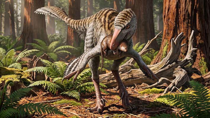
New Troodontid Dinosaur Genus Identified from Fossil Bone Found Decades Ago in New Mexico
Un nouveau genre de dinosaure troodontidé identifié à partir d'un os fossile découvert il y a plusieurs décennies au Nouveau-Mexique
os Mexique fossile Dinevenator Dinosauria Troodontidae oiseau
Les paléontologues ont décrit un nouveau genre de petit dinosaure ressemblant à un oiseau qui vivait dans ce qui est aujourd'hui le Nouveau-Mexique il y a environ 73 millions d'années. L'article Nouveau genre de dinosaures troodontidés identifié à partir d'os fossiles trouvés il y a des décennies au Nouveau-Mexique est apparu en premier sur Sci.News : Breaking Science News.
07/08/2026 sci-news ⚙ Traduction automatique
Le T. rex a-t-il laissé ces marques de morsure vieilles de 66 millions d'années ?
os dent alimentation Dinosauria Tyrannosaurus comportement datation
Les os fossilisés de dinosaures du Wyoming offrent un rare aperçu du comportement alimentaire du Tyrannosaurus rex. Les chercheurs ont examiné plus de 3 000 os datant de 72 à 66 millions d’années et n’en ont trouvé que 12 avec d’éventuelles traces de dents, dont quatre avec des motifs qui correspondaient étroitement à la forme et à l’espacement des dents du T. rex.
05/08/2026 sciencedaily ⚙ Traduction automatique
Célébration de Peter Trusler Palaeoart – Redonner vie aux océans anciens
os fossile Ichthyosauria découverte
L’un des points forts du reportage sur les nouvelles découvertes scientifiques est de voir les œuvres d’art remarquables qui accompagnent de nombreux articles de recherche. Bien que les fossiles en fournissent la preuve, il faut souvent l'habileté d'un paléoartiste talentueux pour transformer les os et les impressions en scènes vivantes des temps profonds. Par exemple, nous avons récemment parlé d’un remarquable fossile d’ichtyosaure.
02/08/2026 everythingdinosaur ⚙ Traduction automatique
Un marteau en os d'éléphant vieux de 500 000 ans montre que les anciens humains étaient des maîtres outilleurs
os découverte
Il y a près de 500 000 ans, les premiers humains d'Angleterre ont transformé un morceau rare d'os d'éléphant en un marteau réutilisable pour aiguiser les outils en pierre. Cette découverte extraordinaire révèle que les anciens outilleurs étaient ingénieux, compétents et capables d’une planification étonnamment avancée.
27/07/2026 sciencedaily ⚙ Traduction automatique
Néandertal semblait manger d’immenses quantités de viande, mais quelque chose ne colle pas
os collagène étude
Des analyses des os de Néandertal révèlent depuis longtemps une énigme alimentaire. Les indices chimiques retrouvés dans leur collagène suggèrent un régime si riche en viande qu'il semble incompatible avec les capacités du corps humain. Une nouvelle étude remet en question une hypothèse...
26/07/2026 futura-terre
1 2 3 4 5 6 7 8 9 10 11