Representative taxa from the Santonian Iharkút fauna from the Csehbánya Formation, Bakony Mountains, western Hungary. A Pannoniasaurus inexpectatus (Squamata, Mosasauroidea), dorsal vertebra (MTM uncatalogued) in dorsal view (photo by Réka Kalmár) B Foxemys trabanti (Pleurodira, Bothremydidae), skull (MTM V 2010.215.1.) in dorsal view (photo by Márton Rabi). C Bicuspidon aff. hatzegiensis (Squamata, Borioteiioidea), left dentary (MTM 2006.112.1.) in medial view (photo by László Makádi) D Basal tetanuran (Theropoda, Tetanurae), tooth (MTM V.01.54) in ?lingual view E Indeterminate abelisaurid (Theropoda, Abelisauridae), pedal ungual phalanx (MTM V 2008.43.1.) in lateral view F Pneumatoraptor fodori (Theropoda, Paraves), left scapulocoracoid (holotype, MTM V 2008.38.1.) in lateral view G Mochlodon vorosi (Ornithopoda, Rhabdodontidae), left dentary (holotype, MTM V 2010.105.1) in lateral view H Bakonydraco galaczi (Pterosauria, Azhdarchidae), mandible (holotype, MTM 2007.110.1) in dorsal view I Iharkutosuchus makadii (Eusuchia, Hylaeochampsidae), skull (holotype, MTM 2006.52.1) in dorsal view J Hungarosaurus tormai (Ankylosauria, Nodosauridae), right dentary (MTM 2007.25.2) in lateral view K Bauxitornis mindszentyae (Aves, Enantiornithes), left tarsometatarsus (holotype, MTM V 2009.38.1) in anterior view L Ajkaceratops kozmai (Ceratopsia), fused rostral and premaxillae (holotype, MTM V 2009.192.1) in lateral view. Scale bars: 2 cm in A, V, G, H, I, J; 1 cm in D, E, F, K, L; 1 mm in C.


![Specimen MN 6117-V, holotype of Oxalaia quilombensis.
A, Left lateral view. B, Right lateral view. C, Dorsal view. D, Slightly oblique ventral view, emphasizing the sculptured condition of the palatal portion of the left premaxilla. Abbreviations for teeth follow Hendrickx et al. [58]. Additional abbreviations: am.p, anteromedial process of maxilla; pm, premaxilla; r.t, replacement tooth; s.p, secondary palate.](https://upload.wikimedia.org/wikipedia/commons/thumb/9/96/Holotype_of_Oxalaia_quilombensis.PNG/330px-Holotype_of_Oxalaia_quilombensis.PNG)


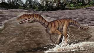
![Reconstruction of the terrestrial paleoenvironmental setting of the Sao Khua Formation by Renata Cunha.
In the center, a generalized spinosaurid feeds on a sauropod. This trophic relationship is hypothesized based on isolated tooth crowns found in association with a sauropod skeleton [67]. In the background, a small pack of the ornithomimosaur theropod Kinnareemimus. Both sauropods and ornithomimosaurs (as part of the “herbivorous” theropods) were found to be positively associated with terrestrial paleoenvironments by Butler and Barrett [15].
(cropped from File:Spinosaurid and Kinnareemimus.PNG)](https://upload.wikimedia.org/wikipedia/commons/thumb/6/62/Kinnareemimus_pack.png/330px-Kinnareemimus_pack.png)