Taxons
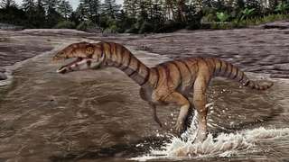
Zby
Cranial and postcranial sauropod remains from sediments of the Praia da Amoreira-Porto Novo Fm. of the coastal sector of Praia da Consolação-Lourinhã-Torres Vedras: 1-2, ?Turiasauria indet., heart-shaped tooth (SHN (JJS) 142, Praia da Corva) in lingual (1) and labial (2) views; 3-4, ?Macronaria indet., spatulate tooth (SHN 513, Porto Novo) in labial (3) and lingual (4) views; 5-6, Macronaria indet., compressed cone-chisel-shaped tooth (SHN 578, Valmitão) in lingual (5) and labial (6) views; 7-8, Eusauropoda indet., partial left maxilla (SHN 582, Praia dos Frades) in lateral (7) and posterior (8) views; 9, Titanosauriformes indet., posterior caudal vertebra (SHN 523, Praia da Corva) in right view; 10-12, Diplodocinae indet., partial skeleton (SHN (JJS) 177, Valmitão), anterior caudal neural spine in posterior view (10), anterior caudal centrum in right view (11) and left ischium in medial view (12); 13-16, holotype material of Zby atlanticus (ML 368, Vale de Pombas), right ungueal I in lateral view (13), right humerus in anterior view (14), right radius in posterior view (15), right ulna in lateral view (16); 17, Eusauropoda indet., partial distal forked-chevron (SHN 587, Praia da Corva) in medial view; 18, Sauropoda indet., pedal ungueal I (SHN 524, Praia de Pedrogãos) in lateral view; 19-22, Macronarian indet., partial skeleton (SHN 181, Valmitão), right astragalus in proximal view (19), anterior caudal vertebra in anterior view (20), right tibia in lateral view (21) and right fibula in medial view (22); 23-24, Eusauropoda indet., partial skeleton (SHN 530, Praia da Corva), anterior chevron in anterior view (23) and anterior caudal vertebra in posterior view (24); 25, cf. Duriatitan humerocristatus, humerus (MG 4976, Praia dos Frades) in anterior view; 26, Sauropoda indet., partial skeleton (SHN 534, Santa Rita), middle chevron in posterior view; 27, Diplodocidae indet., partial skeleton (SHN (JJS) 179, Praia Vermelha), dorsal/caudal (?) neural spine in posterior view; 28-30, holotype material of Dinheirosaurus lourinhanensis (ML 414, Porto Dinheiro), proximal end of a dorsal rib in anterior view (28), anterior caudal neural spine in posterior view (29) and articulated dorsal vertebrae in right view (30). Black scale bar: 10 cm; Grey scale bar: 5 cm; Brown scale bar: 1 cm. See Anatomical abbreviations for abbreviations.


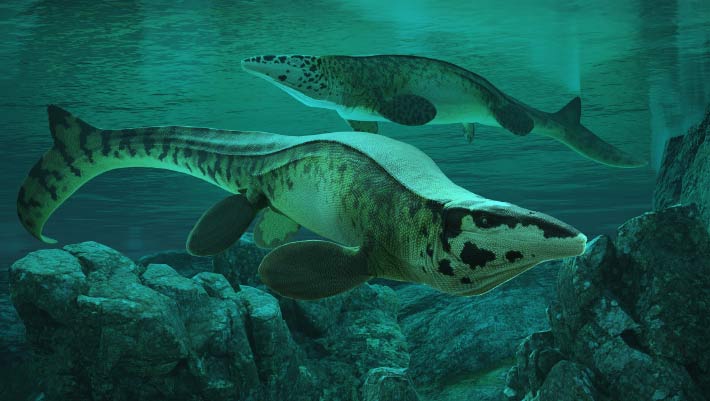
![Partial skull of Shastasaurus pacificus (UCMP 9017) from the Late Triassic of California, USA, in (A) lateral, (B) dorsal, and (C) anterolateral view.
Based on this skull, Shastasaurus has repeatedly been reconstructed with a long, tooth-bearing rostrum. However, note the slenderness of the lower jaw (B, C) and the strong anterior taper of the snout (B), both of which are more consistent with the abbreviated and toothless snout of Shastasaurus liangae comb. nov. than with the traditional long-snouted reconstruction of this skull (as, e.g., in references [22] and [23]).](https://upload.wikimedia.org/wikipedia/commons/thumb/1/1d/Shastasaurus_pacificus.jpg/330px-Shastasaurus_pacificus.jpg)