squelette

Type de vue

294 image(s) · 23 Actualités

Galerie d'images

Holotype specimen of Linheraptor exquisitus, the blocks including nearly the entire skeleton.

Holotype specimen of Linheraptor exquisitus, the blocks including nearly the entire skeleton.

Japon holotype spécimen Linheraptor +1
Hypnovenator matsubaraetoheorum gen. et sp. nov. Blocks including the forelimb, knee (A), and heel (B). (C) Reconstructed skeleton. Red and yellow colors show the confirmed and probable positions of the recovered elements, respectively (Courtesy of Genya Masukawa). (D) Left manus in medial view. (E) Left manual phalanx I-1 in dorsal view. (F) Removed fragmentary left manual phalanx II-3 (manual ungual phalanx II) for preparing the left manus. (G) Cross-section of the bent right ankle. (H) Left metatarsus in posterior view. Abbreviations: as, astragalus; dp, depression; fe, femur; fi, fibula; hu, humerus; mc I, metacarpal I; mc II, metacarpal II; mc III, metacarpal III; mp I-1, manual phalanx I-1; mp I-2, manual phalanx I-2 (manual ungual phalanx I); mp II-1, manual phalanx II-1; mp II-3, manual phalanx II-3 (manual ungual phalanx II); mp III-3, manual phalanx III-3; mp III-4, manual phalanx III-4 (manual ungual phalanx III); mr, medial ridge; mt II, metatarsal II; mt III, metatarsal III; mt IV, metatarsal IV; mt V, metatarsal V; ra, radius; ti, tibia; ul, ulna. Almost all elements are from the left side. Abbreviations for elements from the right side added ‘(r)’ at the end. This figure was created using Adobe Photoshop 25.5.1 and Adobe Illustrator 28.3 (https://www.adobe.com/).

Hypnovenator matsubaraetoheorum gen. et sp. nov. Blocks including the forelimb, knee (A), and heel (B). (C) Reconstructed skeleton. Red and yellow colors show the confirmed and probable positions of the recovered elements, respectively (Courtesy of Genya Masukawa). (D) Left manus in medial view. (E) Left manual phalanx I-1 in dorsal view. (F) Removed fragmentary left manual phalanx II-3 (manual ungual phalanx II) for preparing the left manus. (G) Cross-section of the bent right ankle. (H) Left metatarsus in posterior view. Abbreviations: as, astragalus; dp, depression; fe, femur; fi, fibula; hu, humerus; mc I, metacarpal I; mc II, metacarpal II; mc III, metacarpal III; mp I-1, manual phalanx I-1; mp I-2, manual phalanx I-2 (manual ungual phalanx I); mp II-1, manual phalanx II-1; mp II-3, manual phalanx II-3 (manual ungual phalanx II); mp III-3, manual phalanx III-3; mp III-4, manual phalanx III-4 (manual ungual phalanx III); mr, medial ridge; mt II, metatarsal II; mt III, metatarsal III; mt IV, metatarsal IV; mt V, metatarsal V; ra, radius; ti, tibia; ul, ulna. Almost all elements are from the left side. Abbreviations for elements from the right side added ‘(r)’ at the end. This figure was created using Adobe Photoshop 25.5.1 and Adobe Illustrator 28.3 (https://www.adobe.com/).

humérus Hypnovenator squelette
(a) Photograph. (b) Highly schematic outline shows general layout of the skeleton (illustrated by Zhaochuang). Scale bar, 5 cm. d, dentary; f, femur; fi, fibula; fr, frontal; lj, lower jaw; or, orbit; ti, tibia.

(a) Photograph. (b) Highly schematic outline shows general layout of the skeleton (illustrated by Zhaochuang). Scale bar, 5 cm. d, dentary; f, femur; fi, fibula; fr, frontal; lj, lower jaw; or, orbit; ti, tibia.

écaille Beibeilong squelette
The whole skeleton of the holotype Tongtianlong limosus gen. et sp. nov. in dorsal view (a) and lateral view (b). Scale bar = 10 cm.

The whole skeleton of the holotype Tongtianlong limosus gen. et sp. nov. in dorsal view (a) and lateral view (b). Scale bar = 10 cm.

écaille holotype Tongtianlong squelette
A photograph of the skeleton of Jianchangosaurus yixianensis gen. et sp. nov. (41HIII-0308A) (A) and line drawing (B).

Abbreviations: at, atlas; ax, axis; cav, caudal vertebra; cv, cervical vertebra; co, coracoid; dc, distal carpal; dv, dorsal vertebra; l., left; f, femur; fi, fibula; fur, furcula; hu, humerus; il, ilium; is, ischium; pu, pubis; ra, radius; sc, scapula; ti, tibia; ul, ulna; mc, metacarpal; mt, metatarsal; mxf, maxillary fenestra; pat, proatlas; r., right; sr, sacral rib; sv, sacral vertebra; ung, ungual. All elements of the skeleton are preserved except the distal half of the caudal vertebrae. Dashed lines of metatarsals indicate areas that have been reconstructed. The middle portion of the neck, from the fourth to ninth cervical vertebrae, and the pedal phalanges have been repositioned. The rest of elements of this specimen are in the original position.

A photograph of the skeleton of Jianchangosaurus yixianensis gen. et sp. nov. (41HIII-0308A) (A) and line drawing (B). Abbreviations: at, atlas; ax, axis; cav, caudal vertebra; cv, cervical vertebra; co, coracoid; dc, distal carpal; dv, dorsal vertebra; l., left; f, femur; fi, fibula; fur, furcula; hu, humerus; il, ilium; is, ischium; pu, pubis; ra, radius; sc, scapula; ti, tibia; ul, ulna; mc, metacarpal; mt, metatarsal; mxf, maxillary fenestra; pat, proatlas; r., right; sr, sacral rib; sv, sacral vertebra; ung, ungual. All elements of the skeleton are preserved except the distal half of the caudal vertebrae. Dashed lines of metatarsals indicate areas that have been reconstructed. The middle portion of the neck, from the fourth to ninth cervical vertebrae, and the pedal phalanges have been repositioned. The rest of elements of this specimen are in the original position.

humérus vertèbre dessin spécimen +2
Squelette partiel de Wulatesaurus.

Squelette partiel de Wulatesaurus.

Japon Bannykus partiel squelette
Alvarezsaurus skeleton restoration, Copenhagen.

Alvarezsaurus skeleton restoration, Copenhagen.

Alvarezsauria squelette
Skeleton outline of Dracoraptor hanigani.

Bones highlighted green for present, orange for external moulds and blue for tentatively identified bones. Many unidentified or uncertain elements have been omitted.

Skeleton outline of Dracoraptor hanigani. Bones highlighted green for present, orange for external moulds and blue for tentatively identified bones. Many unidentified or uncertain elements have been omitted.

os Dracoraptor squelette
Reconstruction of the terrestrial paleoenvironmental setting of the Sao Khua Formation by Renata Cunha.
In the center, a generalized spinosaurid feeds on a sauropod. This trophic relationship is hypothesized based on isolated tooth crowns found in association with a sauropod skeleton [67]. In the background, a small pack of the ornithomimosaur theropod Kinnareemimus. Both sauropods and ornithomimosaurs (as part of the “herbivorous” theropods) were found to be positively associated with terrestrial paleoenvironments by Butler and Barrett [15].

(cropped from File:Spinosaurid and Kinnareemimus.PNG)

Reconstruction of the terrestrial paleoenvironmental setting of the Sao Khua Formation by Renata Cunha. In the center, a generalized spinosaurid feeds on a sauropod. This trophic relationship is hypothesized based on isolated tooth crowns found in association with a sauropod skeleton [67]. In the background, a small pack of the ornithomimosaur theropod Kinnareemimus. Both sauropods and ornithomimosaurs (as part of the “herbivorous” theropods) were found to be positively associated with terrestrial paleoenvironments by Butler and Barrett [15]. (cropped from File:Spinosaurid and Kinnareemimus.PNG)

dent Sao Khua Kinnareemimus Ornithomimosauria +3
Photo of the partial holotype skeleton of the ornithomimosaur Garudimimus brevipes, as mounted in the Mongolian Natural History Museum. Scan of physical photograph.

Photo of the partial holotype skeleton of the ornithomimosaur Garudimimus brevipes, as mounted in the Mongolian Natural History Museum. Scan of physical photograph.

musée holotype Garudimimus Ornithomimosauria +2
Reconstruction of the Bravoceratops skeleton with known bones marked in white. Specimen: TMM 46015-1 (holotype). Scale bar=2 m. Total length along centra=6.97 m.

Reconstruction of the Bravoceratops skeleton with known bones marked in white. Specimen: TMM 46015-1 (holotype). Scale bar=2 m. Total length along centra=6.97 m.

os écaille holotype spécimen +2
Ornithischian dinosaur Stenopelix valdensis Meyer, 1857, holotype (GZG 741/2, formerly GPI Gö 741−2), from the Obernkirchen Sandstone (Early
Cretaceous: Berriasian), near Bückeburg, Niedersachsen, Germany. A. Large sandstone slab. B. Interpretative outline drawing of large latex cast (prepared
from large sandstone slab), showing majority of postcranial skeleton in dorsal view. C. Small sandstone slab. D. Interpretative outline drawing of small la−
tex cast (prepared from small sandstone slab), showing sacrum and caudals, pelvic region and partial hindlimbs in ventral view. For clarity elements in and
around the sacral region have not been labelled—these areas are shown in greater detail in Fig. 3. Roman numerals II–IV correspond to respective digits.

Abbreviations: mt, metatarsals; f, femur; ?, unidentified element.

Ornithischian dinosaur Stenopelix valdensis Meyer, 1857, holotype (GZG 741/2, formerly GPI Gö 741−2), from the Obernkirchen Sandstone (Early Cretaceous: Berriasian), near Bückeburg, Niedersachsen, Germany. A. Large sandstone slab. B. Interpretative outline drawing of large latex cast (prepared from large sandstone slab), showing majority of postcranial skeleton in dorsal view. C. Small sandstone slab. D. Interpretative outline drawing of small la− tex cast (prepared from small sandstone slab), showing sacrum and caudals, pelvic region and partial hindlimbs in ventral view. For clarity elements in and around the sacral region have not been labelled—these areas are shown in greater detail in Fig. 3. Roman numerals II–IV correspond to respective digits. Abbreviations: mt, metatarsals; f, femur; ?, unidentified element.

dessin Allemagne Berriasien Crétacé +8
Composite skeleton on display at the Perot Museum of Nature and Science. Scale bar equals 10 cm.

Composite skeleton on display at the Perot Museum of Nature and Science. Scale bar equals 10 cm.

écaille musée Convolosaurus squelette
The holotype skeleton of Kamuysaurus japonicus gen. et sp. nov. (b) Reconstructed skeleton, showing recovered elements. Selected postcranial elements: cervical vertebrae (atlas, axis, and fourth and twelfth cervicals) in left lateral view (c), dorsal vertebrae (first, seventh, and sixteenth dorsals) in left lateral view (d), caudal vertebrae (anterior, middle, and posterior caudals) in left lateral view (e), left scapula (f) and coracoid (g) in lateral view, right sternum in ventral view (h), left humerus in anterior view (i), right ulna and radius in medial view (j), right manus in dorsal view (k), right pelvis in lateral view (l), right femur in anterior view (m), right tibia in anterior view (n), right fibula in lateral view (o), right astragalus and calcaneum, articulated positioned with tibia (p), and right pes in dorsal view (q). All scales are 10 cm except 1 m scale for (b). Abbreviations; ac, acromion process; ast, astragalus; cal, calcaneum; dpc, deltopectoral crest; fi, fibula; icg, intercondylar groove; il, ilium; is, ischium; ltr, lesser trochanter; mc2, metacarpal II; mc5, metacarpal V; mt2, metatarsal II; mt4, metatarsal IV; nc, neurocentrum; od, odontoid; olp, olecranon process; pc, pleurocentrum; pu, pubis; ra, radius; ti, tibia; ul, ulna; vp, ventral process.

The holotype skeleton of Kamuysaurus japonicus gen. et sp. nov. (b) Reconstructed skeleton, showing recovered elements. Selected postcranial elements: cervical vertebrae (atlas, axis, and fourth and twelfth cervicals) in left lateral view (c), dorsal vertebrae (first, seventh, and sixteenth dorsals) in left lateral view (d), caudal vertebrae (anterior, middle, and posterior caudals) in left lateral view (e), left scapula (f) and coracoid (g) in lateral view, right sternum in ventral view (h), left humerus in anterior view (i), right ulna and radius in medial view (j), right manus in dorsal view (k), right pelvis in lateral view (l), right femur in anterior view (m), right tibia in anterior view (n), right fibula in lateral view (o), right astragalus and calcaneum, articulated positioned with tibia (p), and right pes in dorsal view (q). All scales are 10 cm except 1 m scale for (b). Abbreviations; ac, acromion process; ast, astragalus; cal, calcaneum; dpc, deltopectoral crest; fi, fibula; icg, intercondylar groove; il, ilium; is, ischium; ltr, lesser trochanter; mc2, metacarpal II; mc5, metacarpal V; mt2, metatarsal II; mt4, metatarsal IV; nc, neurocentrum; od, odontoid; olp, olecranon process; pc, pleurocentrum; pu, pubis; ra, radius; ti, tibia; ul, ulna; vp, ventral process.

crête humérus bassin écaille +3
Incomplete skeleton of Camptosaurus prestwichii, now referred to as Cumnoria, Oxford University Museum of Natural History.

Incomplete skeleton of Camptosaurus prestwichii, now referred to as Cumnoria, Oxford University Museum of Natural History.

musée Camptosaurus Cumnoria squelette
Yueosaurus tiantaiensis, reconstructed skeletons, Zhejiang Geological Museum

Yueosaurus tiantaiensis, reconstructed skeletons, Zhejiang Geological Museum

musée Yueosaurus squelette
1 2 3 4 5 6 7 8 9 10 11 12 13 14 15 16 17 18 19

Actualités

Fossil of Giant Squid-Like Creature from Cretaceous Period Bears Marks of Deadly Attack
Canada Crétacé fossile squelette
Paleontologists studying a fossil unearthed in southern Manitoba, Canada, have identified what they believe are two bite marks on the internal skeleton of the ancient giant cephalopod Enchoteuthis. The post Fossil of Giant Squid-Like Creature from Cretaceous Period Bears Marks of Deadly Attack appeared first on Sci.News: Breaking Science News.
01/09/2026 sci-news
Un remarquable fossile de reptile marin du Trias préserve les organes internes
Chine Trias fossile spécimen anatomie squelette
Les scientifiques ont décrit un fossile exceptionnel du reptile marin du Trias au long cou, Austronaga minuta. Le spécimen vient de Chine et a environ 245 millions d’années. Remarquablement, une grande partie du système digestif de l’animal a été préservée. Le fossile presque complet offre aux scientifiques une vue sans précédent du squelette et de l’anatomie interne d’un archosauromorphe.
26/08/2026 everythingdinosaur ⚙ Traduction automatique
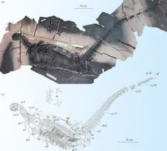
Un fossile remarquable révèle le régime alimentaire d'un ichtyosaure du Jurassique supérieur
Allemagne Jurassique Jurassique supérieur fossile spécimen Aegirosaurus Ichthyosauria Jabalisaurus squelette
Les scientifiques étudiant l’ichtyosaure Jabalisaurus tethyensis, récemment décrit, ont acquis un aperçu remarquable de son régime alimentaire. Un fossile magnifiquement conservé du sud de l'Allemagne contient des preuves des derniers repas de l'animal. De plus, le spécimen préserve des détails importants du squelette et des contours du corps. Le fossile, numéro de spécimen JME-SOS-08369, était auparavant attribué à Aegirosaurus. Cependant, les chercheurs
24/08/2026 everythingdinosaur ⚙ Traduction automatique
Spoon-Toothed, Short-Headed Marine Reptile Unearthed in China
Un reptile marin à dents de cuillère et à tête courte découvert en Chine
dent Chine Trias Sauropterygia nouvelle espèce autres reptiles squelette
Un nouveau genre et une nouvelle espèce de reptile sauroptérygien ayant vécu pendant la période du Trias, il y a environ 245 millions d'années, ont été identifiés à partir d'un squelette presque complet trouvé en Chine. L'article Un reptile marin à dents de cuillère et à tête courte découvert en Chine est apparu en premier sur Sci.News : Breaking Science News.
20/08/2026 sci-news ⚙ Traduction automatique
Des squelettes vieux de 3 500 ans remettent en question ce que les scientifiques pensaient savoir sur la syphilis
Viêt Nam découverte squelette
Des vestiges anciens du Vietnam suggèrent que des maladies comme le pian pourraient avoir été transmises de la mère à l'enfant il y a des milliers d'années, une forme de transmission souvent supposée indiquer la syphilis. Cette découverte pourrait bouleverser la façon dont les scientifiques interprètent les infections anciennes et compliquer le mystère de longue date des origines de la syphilis.
12/08/2026 sciencedaily-paleo ⚙ Traduction automatique
1 2 3 4 5