squelette

Type de vue

136 image(s) · 8 Actualités

Galerie d'images

Entire skeleton of Jidapterus edentus (RCPS-030366CY) preserved in a slab mostly in right lateral view.

Abbreviations: aa, the atlas-axis complex; bo, basioccipital; br, braincase; cve, cervical vertebrae; cv3, the third cervical vertebra; cv8, the eighth cervical vertebra; ds, dentaries; dv, dorsal vertebra; dve, dorsal vertebrae; ep, epiphysis; j, jugal; lc, left carpus; lf, left femur; li, left ilium; lis, left ischium; lm, left manual digits; lp, left pubis; lpe, left pes; lpt, left pteroid; lr, left radius; lsc, left scapula-coracoid; lt, left tibia; lta, left tarsus; lu, left ulna; lwmc, left wing metacarpal; lwp1–4; left wing phalanges 1–4; p, pterygoid; pp, postacetabular process of the right ilium; q, quadrate; rc, right carpus; rh, right humerus; rf, right femur; rfi, right fibula; ribs, ribs; ris, right ischium; rm, right manual digits; rmcI, right metacarpal I; rp, right pubis; rpe, right pes; rpt, right pteroid; rr, right radius; rsc, right scapula-coracoid; rt, right tibia; ru, right ulna; rwmc, right wing metacarpal; rwp1–4, right wing phalanges 1–4; rostrum, rostrum; st, sternum; sv, sacral vertebra; sve, sacral vertebrae. Gray shaded areas bound by solid lines indicate preserved bones, dashed lines indicate bones with margins discerned by impression, and lack of lines indicate missing bone or displacement along cracks in the slab.
Taxons Jidapterus

Entire skeleton of Jidapterus edentus (RCPS-030366CY) preserved in a slab mostly in right lateral view. Abbreviations: aa, the atlas-axis complex; bo, basioccipital; br, braincase; cve, cervical vertebrae; cv3, the third cervical vertebra; cv8, the eighth cervical vertebra; ds, dentaries; dv, dorsal vertebra; dve, dorsal vertebrae; ep, epiphysis; j, jugal; lc, left carpus; lf, left femur; li, left ilium; lis, left ischium; lm, left manual digits; lp, left pubis; lpe, left pes; lpt, left pteroid; lr, left radius; lsc, left scapula-coracoid; lt, left tibia; lta, left tarsus; lu, left ulna; lwmc, left wing metacarpal; lwp1–4; left wing phalanges 1–4; p, pterygoid; pp, postacetabular process of the right ilium; q, quadrate; rc, right carpus; rh, right humerus; rf, right femur; rfi, right fibula; ribs, ribs; ris, right ischium; rm, right manual digits; rmcI, right metacarpal I; rp, right pubis; rpe, right pes; rpt, right pteroid; rr, right radius; rsc, right scapula-coracoid; rt, right tibia; ru, right ulna; rwmc, right wing metacarpal; rwp1–4, right wing phalanges 1–4; rostrum, rostrum; st, sternum; sv, sacral vertebra; sve, sacral vertebrae. Gray shaded areas bound by solid lines indicate preserved bones, dashed lines indicate bones with margins discerned by impression, and lack of lines indicate missing bone or displacement along cracks in the slab.

humérus vertèbre Chaoyangopteridae Chaoyangopterinae +2
Entire skeleton of Jidapterus edentus (RCPS-030366CY) preserved in a slab mostly in right lateral view.

Abbreviations: aa, the atlas-axis complex; bo, basioccipital; br, braincase; cve, cervical vertebrae; cv3, the third cervical vertebra; cv8, the eighth cervical vertebra; ds, dentaries; dv, dorsal vertebra; dve, dorsal vertebrae; ep, epiphysis; j, jugal; lc, left carpus; lf, left femur; li, left ilium; lis, left ischium; lm, left manual digits; lp, left pubis; lpe, left pes; lpt, left pteroid; lr, left radius; lsc, left scapula-coracoid; lt, left tibia; lta, left tarsus; lu, left ulna; lwmc, left wing metacarpal; lwp1–4; left wing phalanges 1–4; p, pterygoid; pp, postacetabular process of the right ilium; q, quadrate; rc, right carpus; rh, right humerus; rf, right femur; rfi, right fibula; ribs, ribs; ris, right ischium; rm, right manual digits; rmcI, right metacarpal I; rp, right pubis; rpe, right pes; rpt, right pteroid; rr, right radius; rsc, right scapula-coracoid; rt, right tibia; ru, right ulna; rwmc, right wing metacarpal; rwp1–4, right wing phalanges 1–4; rostrum, rostrum; st, sternum; sv, sacral vertebra; sve, sacral vertebrae. Gray shaded areas bound by solid lines indicate preserved bones, dashed lines indicate bones with margins discerned by impression, and lack of lines indicate missing bone or displacement along cracks in the slab.
Taxons Chaoyangopteridae

Entire skeleton of Jidapterus edentus (RCPS-030366CY) preserved in a slab mostly in right lateral view. Abbreviations: aa, the atlas-axis complex; bo, basioccipital; br, braincase; cve, cervical vertebrae; cv3, the third cervical vertebra; cv8, the eighth cervical vertebra; ds, dentaries; dv, dorsal vertebra; dve, dorsal vertebrae; ep, epiphysis; j, jugal; lc, left carpus; lf, left femur; li, left ilium; lis, left ischium; lm, left manual digits; lp, left pubis; lpe, left pes; lpt, left pteroid; lr, left radius; lsc, left scapula-coracoid; lt, left tibia; lta, left tarsus; lu, left ulna; lwmc, left wing metacarpal; lwp1–4; left wing phalanges 1–4; p, pterygoid; pp, postacetabular process of the right ilium; q, quadrate; rc, right carpus; rh, right humerus; rf, right femur; rfi, right fibula; ribs, ribs; ris, right ischium; rm, right manual digits; rmcI, right metacarpal I; rp, right pubis; rpe, right pes; rpt, right pteroid; rr, right radius; rsc, right scapula-coracoid; rt, right tibia; ru, right ulna; rwmc, right wing metacarpal; rwp1–4, right wing phalanges 1–4; rostrum, rostrum; st, sternum; sv, sacral vertebra; sve, sacral vertebrae. Gray shaded areas bound by solid lines indicate preserved bones, dashed lines indicate bones with margins discerned by impression, and lack of lines indicate missing bone or displacement along cracks in the slab.

humérus vertèbre Chaoyangopteridae Chaoyangopterinae +2
Entire skeleton of Jidapterus edentus (RCPS-030366CY) preserved in a slab mostly in right lateral view.

Abbreviations: aa, the atlas-axis complex; bo, basioccipital; br, braincase; cve, cervical vertebrae; cv3, the third cervical vertebra; cv8, the eighth cervical vertebra; ds, dentaries; dv, dorsal vertebra; dve, dorsal vertebrae; ep, epiphysis; j, jugal; lc, left carpus; lf, left femur; li, left ilium; lis, left ischium; lm, left manual digits; lp, left pubis; lpe, left pes; lpt, left pteroid; lr, left radius; lsc, left scapula-coracoid; lt, left tibia; lta, left tarsus; lu, left ulna; lwmc, left wing metacarpal; lwp1–4; left wing phalanges 1–4; p, pterygoid; pp, postacetabular process of the right ilium; q, quadrate; rc, right carpus; rh, right humerus; rf, right femur; rfi, right fibula; ribs, ribs; ris, right ischium; rm, right manual digits; rmcI, right metacarpal I; rp, right pubis; rpe, right pes; rpt, right pteroid; rr, right radius; rsc, right scapula-coracoid; rt, right tibia; ru, right ulna; rwmc, right wing metacarpal; rwp1–4, right wing phalanges 1–4; rostrum, rostrum; st, sternum; sv, sacral vertebra; sve, sacral vertebrae. Gray shaded areas bound by solid lines indicate preserved bones, dashed lines indicate bones with margins discerned by impression, and lack of lines indicate missing bone or displacement along cracks in the slab.
Taxons Chaoyangopterinae

Entire skeleton of Jidapterus edentus (RCPS-030366CY) preserved in a slab mostly in right lateral view. Abbreviations: aa, the atlas-axis complex; bo, basioccipital; br, braincase; cve, cervical vertebrae; cv3, the third cervical vertebra; cv8, the eighth cervical vertebra; ds, dentaries; dv, dorsal vertebra; dve, dorsal vertebrae; ep, epiphysis; j, jugal; lc, left carpus; lf, left femur; li, left ilium; lis, left ischium; lm, left manual digits; lp, left pubis; lpe, left pes; lpt, left pteroid; lr, left radius; lsc, left scapula-coracoid; lt, left tibia; lta, left tarsus; lu, left ulna; lwmc, left wing metacarpal; lwp1–4; left wing phalanges 1–4; p, pterygoid; pp, postacetabular process of the right ilium; q, quadrate; rc, right carpus; rh, right humerus; rf, right femur; rfi, right fibula; ribs, ribs; ris, right ischium; rm, right manual digits; rmcI, right metacarpal I; rp, right pubis; rpe, right pes; rpt, right pteroid; rr, right radius; rsc, right scapula-coracoid; rt, right tibia; ru, right ulna; rwmc, right wing metacarpal; rwp1–4, right wing phalanges 1–4; rostrum, rostrum; st, sternum; sv, sacral vertebra; sve, sacral vertebrae. Gray shaded areas bound by solid lines indicate preserved bones, dashed lines indicate bones with margins discerned by impression, and lack of lines indicate missing bone or displacement along cracks in the slab.

humérus vertèbre Chaoyangopteridae Chaoyangopterinae +2
Reconstructed Thalassodromeus sethi skeleton (the postcranium is hypothetical) with Anhanguera behind, National Museum of Nature and Science, Tokyo.
Taxons Thalassodromeus

Reconstructed Thalassodromeus sethi skeleton (the postcranium is hypothetical) with Anhanguera behind, National Museum of Nature and Science, Tokyo.

musée Anhanguera Thalassodromeus squelette
Nemicolopterus crypticus holotype (IVPP V-14377).

(A) Skeleton overview, and (B) schematic drawing. (C) Skull (right lateral view), and (D) schematic drawing. Abbreviations: cdv, caudal vertebrae; co, coracoid; cv, cervical vertebra; d1–d4, digits 1–4; f, frontal; fe, femur; h, humerus; hy, hyoid; il, illium; is, ischium; j, jugal; l, left; la, lacrimal; mand, mandible; mc, metacarpal; mt, metatarsal; naof, nasoantorbital fenestra; or, orbit; pa, parietal; pm, premaxilla; ph, phalanx; ti, tibia; r, right; sca, scapula. Scale bars: A–B, 100 mm; C–D, 5 mm.
Taxons Nemicolopterus

Nemicolopterus crypticus holotype (IVPP V-14377). (A) Skeleton overview, and (B) schematic drawing. (C) Skull (right lateral view), and (D) schematic drawing. Abbreviations: cdv, caudal vertebrae; co, coracoid; cv, cervical vertebra; d1–d4, digits 1–4; f, frontal; fe, femur; h, humerus; hy, hyoid; il, illium; is, ischium; j, jugal; l, left; la, lacrimal; mand, mandible; mc, metacarpal; mt, metatarsal; naof, nasoantorbital fenestra; or, orbit; pa, parietal; pm, premaxilla; ph, phalanx; ti, tibia; r, right; sca, scapula. Scale bars: A–B, 100 mm; C–D, 5 mm.

humérus écaille vertèbre dessin +4
Selected elements used in the diagnosis of Saltriovenator zanellai n. gen. n. sp.

Right humerus in medial (A), frontal (B) and distal (C) views; (D) left scapula, medial view; (E) right scapular glenoid and coracoid, lateral view; (F) furcula, ventral view; tooth, labial (G) and apical (H) views; (I) left humerus, medial view; right second metacarpal in dorsal (J), lateral (L) and distal (N) views; first phalanx of the right second digit in dorsal (K), lateral (M) and proximal (O) views; (P–T) right third digit in proximal, dorsal and lateral views; (U) right distal tarsal IV, proximal view; third right metatarsal in proximal (V) and frontal (X) views; second right metatarsal, proximal (W) and frontal (Y) views; (Z) reconstructed skeleton showing identified elements (red). Abbreviations as in text, asterisks mark autapomorphic traits. Scale bars: 10 cm in (A)–(E), (I), and (U)–(Y); two cm in (F), and (J)–(T); one cm in (G).
Taxons Saltriovenator

Selected elements used in the diagnosis of Saltriovenator zanellai n. gen. n. sp. Right humerus in medial (A), frontal (B) and distal (C) views; (D) left scapula, medial view; (E) right scapular glenoid and coracoid, lateral view; (F) furcula, ventral view; tooth, labial (G) and apical (H) views; (I) left humerus, medial view; right second metacarpal in dorsal (J), lateral (L) and distal (N) views; first phalanx of the right second digit in dorsal (K), lateral (M) and proximal (O) views; (P–T) right third digit in proximal, dorsal and lateral views; (U) right distal tarsal IV, proximal view; third right metatarsal in proximal (V) and frontal (X) views; second right metatarsal, proximal (W) and frontal (Y) views; (Z) reconstructed skeleton showing identified elements (red). Abbreviations as in text, asterisks mark autapomorphic traits. Scale bars: 10 cm in (A)–(E), (I), and (U)–(Y); two cm in (F), and (J)–(T); one cm in (G).

humérus écaille dent Saltriovenator +1
Eustreptospondylus skeleton
Taxons Eustreptospondylus

Eustreptospondylus skeleton

Eustreptospondylus squelette
Holotype specimen of Linheraptor exquisitus, the blocks including nearly the entire skeleton.
Taxons Linheraptor

Holotype specimen of Linheraptor exquisitus, the blocks including nearly the entire skeleton.

holotype spécimen Linheraptor squelette
Hypnovenator matsubaraetoheorum gen. et sp. nov. Blocks including the forelimb, knee (A), and heel (B). (C) Reconstructed skeleton. Red and yellow colors show the confirmed and probable positions of the recovered elements, respectively (Courtesy of Genya Masukawa). (D) Left manus in medial view. (E) Left manual phalanx I-1 in dorsal view. (F) Removed fragmentary left manual phalanx II-3 (manual ungual phalanx II) for preparing the left manus. (G) Cross-section of the bent right ankle. (H) Left metatarsus in posterior view. Abbreviations: as, astragalus; dp, depression; fe, femur; fi, fibula; hu, humerus; mc I, metacarpal I; mc II, metacarpal II; mc III, metacarpal III; mp I-1, manual phalanx I-1; mp I-2, manual phalanx I-2 (manual ungual phalanx I); mp II-1, manual phalanx II-1; mp II-3, manual phalanx II-3 (manual ungual phalanx II); mp III-3, manual phalanx III-3; mp III-4, manual phalanx III-4 (manual ungual phalanx III); mr, medial ridge; mt II, metatarsal II; mt III, metatarsal III; mt IV, metatarsal IV; mt V, metatarsal V; ra, radius; ti, tibia; ul, ulna. Almost all elements are from the left side. Abbreviations for elements from the right side added ‘(r)’ at the end. This figure was created using Adobe Photoshop 25.5.1 and Adobe Illustrator 28.3 (https://www.adobe.com/).
Taxons Hypnovenator

Hypnovenator matsubaraetoheorum gen. et sp. nov. Blocks including the forelimb, knee (A), and heel (B). (C) Reconstructed skeleton. Red and yellow colors show the confirmed and probable positions of the recovered elements, respectively (Courtesy of Genya Masukawa). (D) Left manus in medial view. (E) Left manual phalanx I-1 in dorsal view. (F) Removed fragmentary left manual phalanx II-3 (manual ungual phalanx II) for preparing the left manus. (G) Cross-section of the bent right ankle. (H) Left metatarsus in posterior view. Abbreviations: as, astragalus; dp, depression; fe, femur; fi, fibula; hu, humerus; mc I, metacarpal I; mc II, metacarpal II; mc III, metacarpal III; mp I-1, manual phalanx I-1; mp I-2, manual phalanx I-2 (manual ungual phalanx I); mp II-1, manual phalanx II-1; mp II-3, manual phalanx II-3 (manual ungual phalanx II); mp III-3, manual phalanx III-3; mp III-4, manual phalanx III-4 (manual ungual phalanx III); mr, medial ridge; mt II, metatarsal II; mt III, metatarsal III; mt IV, metatarsal IV; mt V, metatarsal V; ra, radius; ti, tibia; ul, ulna. Almost all elements are from the left side. Abbreviations for elements from the right side added ‘(r)’ at the end. This figure was created using Adobe Photoshop 25.5.1 and Adobe Illustrator 28.3 (https://www.adobe.com/).

humérus Hypnovenator squelette
(a) Photograph. (b) Highly schematic outline shows general layout of the skeleton (illustrated by Zhaochuang). Scale bar, 5 cm. d, dentary; f, femur; fi, fibula; fr, frontal; lj, lower jaw; or, orbit; ti, tibia.
Taxons Beibeilong

(a) Photograph. (b) Highly schematic outline shows general layout of the skeleton (illustrated by Zhaochuang). Scale bar, 5 cm. d, dentary; f, femur; fi, fibula; fr, frontal; lj, lower jaw; or, orbit; ti, tibia.

écaille Beibeilong squelette
The whole skeleton of the holotype Tongtianlong limosus gen. et sp. nov. in dorsal view (a) and lateral view (b). Scale bar = 10 cm.
Taxons Tongtianlong

The whole skeleton of the holotype Tongtianlong limosus gen. et sp. nov. in dorsal view (a) and lateral view (b). Scale bar = 10 cm.

écaille holotype Tongtianlong squelette
A photograph of the skeleton of Jianchangosaurus yixianensis gen. et sp. nov. (41HIII-0308A) (A) and line drawing (B).

Abbreviations: at, atlas; ax, axis; cav, caudal vertebra; cv, cervical vertebra; co, coracoid; dc, distal carpal; dv, dorsal vertebra; l., left; f, femur; fi, fibula; fur, furcula; hu, humerus; il, ilium; is, ischium; pu, pubis; ra, radius; sc, scapula; ti, tibia; ul, ulna; mc, metacarpal; mt, metatarsal; mxf, maxillary fenestra; pat, proatlas; r., right; sr, sacral rib; sv, sacral vertebra; ung, ungual. All elements of the skeleton are preserved except the distal half of the caudal vertebrae. Dashed lines of metatarsals indicate areas that have been reconstructed. The middle portion of the neck, from the fourth to ninth cervical vertebrae, and the pedal phalanges have been repositioned. The rest of elements of this specimen are in the original position.
Taxons Jianchangosaurus

A photograph of the skeleton of Jianchangosaurus yixianensis gen. et sp. nov. (41HIII-0308A) (A) and line drawing (B). Abbreviations: at, atlas; ax, axis; cav, caudal vertebra; cv, cervical vertebra; co, coracoid; dc, distal carpal; dv, dorsal vertebra; l., left; f, femur; fi, fibula; fur, furcula; hu, humerus; il, ilium; is, ischium; pu, pubis; ra, radius; sc, scapula; ti, tibia; ul, ulna; mc, metacarpal; mt, metatarsal; mxf, maxillary fenestra; pat, proatlas; r., right; sr, sacral rib; sv, sacral vertebra; ung, ungual. All elements of the skeleton are preserved except the distal half of the caudal vertebrae. Dashed lines of metatarsals indicate areas that have been reconstructed. The middle portion of the neck, from the fourth to ninth cervical vertebrae, and the pedal phalanges have been repositioned. The rest of elements of this specimen are in the original position.

humérus vertèbre dessin spécimen +2
Squelette partiel de Wulatesaurus.
Taxons Bannykus

Squelette partiel de Wulatesaurus.

Bannykus partiel squelette
Alvarezsaurus skeleton restoration, Copenhagen.
Taxons Alvarezsaurus

Alvarezsaurus skeleton restoration, Copenhagen.

restauration Alvarezsaurus squelette
Skeleton outline of Dracoraptor hanigani.

Bones highlighted green for present, orange for external moulds and blue for tentatively identified bones. Many unidentified or uncertain elements have been omitted.
Taxons Dracoraptor

Skeleton outline of Dracoraptor hanigani. Bones highlighted green for present, orange for external moulds and blue for tentatively identified bones. Many unidentified or uncertain elements have been omitted.

Dracoraptor squelette
Reconstruction of the terrestrial paleoenvironmental setting of the Sao Khua Formation by Renata Cunha.
In the center, a generalized spinosaurid feeds on a sauropod. This trophic relationship is hypothesized based on isolated tooth crowns found in association with a sauropod skeleton [67]. In the background, a small pack of the ornithomimosaur theropod Kinnareemimus. Both sauropods and ornithomimosaurs (as part of the “herbivorous” theropods) were found to be positively associated with terrestrial paleoenvironments by Butler and Barrett [15].

(cropped from File:Spinosaurid and Kinnareemimus.PNG)
Taxons Kinnareemimus

Reconstruction of the terrestrial paleoenvironmental setting of the Sao Khua Formation by Renata Cunha. In the center, a generalized spinosaurid feeds on a sauropod. This trophic relationship is hypothesized based on isolated tooth crowns found in association with a sauropod skeleton [67]. In the background, a small pack of the ornithomimosaur theropod Kinnareemimus. Both sauropods and ornithomimosaurs (as part of the “herbivorous” theropods) were found to be positively associated with terrestrial paleoenvironments by Butler and Barrett [15]. (cropped from File:Spinosaurid and Kinnareemimus.PNG)

dent reconstitution Sao Khua Kinnareemimus +3
1 2 3 4 5 6 7 8 9

Actualités

Ce dinosaure de 2 livres réécrit ce que les scientifiques savent de l'évolution
fossile Alnashetri Alvarezsaurus Dinosaurus oiseau évolution squelette
Un squelette de dinosaure presque complet découvert en Patagonie aide les scientifiques à percer le mystère des alvarezsaures, un groupe étrange de dinosaures ressemblant à des oiseaux. Le fossile d'Alnashetri cerropoliciensis révèle que ces animaux sont devenus minuscules avant de développer leurs caractéristiques spécialisées ultérieures, telles que des bras tronqués et des adaptations mangeuses de fourmis. Pesant moins de deux livres, le dinosaure est l’un des plus petits connus d’Amérique du Sud.
10/03/2026 sciencedaily ⚙ Traduction automatique
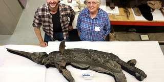
Un fossile de « Sword Dragon » vieux de 190 millions d’années réécrit l’histoire des ichtyosaures
Royaume-Uni Jurassique fossile évolution squelette
Un ichtyosaure nouvellement identifié de la côte jurassique du Royaume-Uni est en train de réécrire une partie du manuel préhistorique. Surnommé le « dragon-épée du Dorset », ce reptile marin de trois mètres de long a vécu pendant une période d’évolution mal comprise, au cours de laquelle les principaux groupes d’ichtyosaures disparaissaient et de nouveaux apparaissaient. Son squelette magnifiquement préservé – doté d’un museau en forme de lame et d’un éventuel dernier repas – permet de déterminer le moment où cette transition dramatique s’est produite.
24/02/2026 sciencedaily ⚙ Traduction automatique
Des os de dinosaures découverts presque les uns sur les autres en Transylvanie
os bassin Roumanie fossile Dinosaurus Titanosaurus squelette
Des scientifiques explorant le bassin de Hațeg en Roumanie ont découvert l’un des sites de fossiles de dinosaures les plus denses jamais découverts, avec des os presque les uns sur les autres. Le site K2 préserve des milliers de vestiges d’un lac préhistorique alimenté en crue qui agissait comme un piège osseux naturel il y a 72 millions d’années. Aux côtés de dinosaures locaux communs, les chercheurs ont découvert les premiers squelettes de titanosaures bien conservés jamais découverts dans la région. Le site révèle comment les anciens écosystèmes européens de dinosaures se sont formés et ont évolué au cours de cette période.
23/12/2025 sciencedaily ⚙ Traduction automatique
1 2