squelette

Type de vue

167 image(s) · 17 Actualités

Galerie d'images

Hypothetical reconstruction of the skeleton of Amanasaurus nesbitti gen. et sp. nov. depicting (in orange) the preserved portions. (a) CAPPA/UFSM 0374 (holotype), a proximal portion of a right femur in anterior view. (b) CAPPA/UFSM 0375 (referred specimen), a distal portion of a left femur in anterior view. Figures were generated with GIMP 2.8 (https://www.gimp.org/).
Cropped from original image and letters adjusted to match.

Hypothetical reconstruction of the skeleton of Amanasaurus nesbitti gen. et sp. nov. depicting (in orange) the preserved portions. (a) CAPPA/UFSM 0374 (holotype), a proximal portion of a right femur in anterior view. (b) CAPPA/UFSM 0375 (referred specimen), a distal portion of a left femur in anterior view. Figures were generated with GIMP 2.8 (https://www.gimp.org/). Cropped from original image and letters adjusted to match.

holotype spécimen Amanasaurus squelette
Pliosaurus (Luskhan itilensis) lived on the territory of the Volga region in the Hauterivian age of the Early Cretaceous period. Discovered in 2002 by G.N. Uspensky on the banks of the Volga near the village of Slantsevy Rudnik. This is the most complete pliosaurus skeleton found in Russia. This pliosaurus was not a predator and preferred to feed on fish and cephalopods.

Pliosaurus (Luskhan itilensis) lived on the territory of the Volga region in the Hauterivian age of the Early Cretaceous period. Discovered in 2002 by G.N. Uspensky on the banks of the Volga near the village of Slantsevy Rudnik. This is the most complete pliosaurus skeleton found in Russia. This pliosaurus was not a predator and preferred to feed on fish and cephalopods.

prédateur Russie Crétacé Crétacé inférieur +5
Skeleton and reconstruction of Luskhan
Taxons Luskhan

Skeleton and reconstruction of Luskhan

Luskhan squelette
Holotype of Alcione elainus.
Fig. 6 of:
Longrich, N. R., Martill, D. M., & Andres, B. (2018). Late Maastrichtian pterosaurs from North Africa and mass extinction of Pterosauria at the Cretaceous-Paleogene boundary. PLoS biology, 16(3), e2001663.
---
Original figure legend:
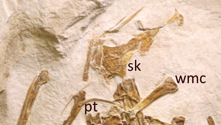
A. elainus FSAC-OB 2, holotype partial skeleton and FSAC-OB 217, metacarpal IV.

(A) Holotype right humerus in anterior view, (B) holotype right ulna and radius in anterior view, respectively, (C) holotype sternum in left lateral view, (D) referred metacarpal IV, (E) holotype, distal end of left metacarpal IV and left scapulocoracoid, and (F) holotype right femur in posterior view. Abbreviations: co, coracoid; cr, cristospine; dc, distal condyle; dpc, deltopectoral crest; ect, ectepicondyle; fh, femoral head; gl, glenoid; gt, greater trochanter; hh, humeral head; hum, humerus; mcIV, metacarpal IV, pc, proximal cotyle; pf, pneumatic foramen; rad, radius; scpr, supracondylar process; ste, sternum; uln, ulna.
Taxons Alcione

Holotype of Alcione elainus. Fig. 6 of: Longrich, N. R., Martill, D. M., & Andres, B. (2018). Late Maastrichtian pterosaurs from North Africa and mass extinction of Pterosauria at the Cretaceous-Paleogene boundary. PLoS biology, 16(3), e2001663. --- Original figure legend: A. elainus FSAC-OB 2, holotype partial skeleton and FSAC-OB 217, metacarpal IV. (A) Holotype right humerus in anterior view, (B) holotype right ulna and radius in anterior view, respectively, (C) holotype sternum in left lateral view, (D) referred metacarpal IV, (E) holotype, distal end of left metacarpal IV and left scapulocoracoid, and (F) holotype right femur in posterior view. Abbreviations: co, coracoid; cr, cristospine; dc, distal condyle; dpc, deltopectoral crest; ect, ectepicondyle; fh, femoral head; gl, glenoid; gt, greater trochanter; hh, humeral head; hum, humerus; mcIV, metacarpal IV, pc, proximal cotyle; pf, pneumatic foramen; rad, radius; scpr, supracondylar process; ste, sternum; uln, ulna.

crête humérus Crétacé Maastrichtien +7
Identifier: annalsofmedicalh01pack (find matches)
Title: Annals of medical history
Year: 1917 (1910s)
Authors:  Packard, Francis R. (Francis Randolph), 1870-1950
Subjects:  Medicine
Publisher:  New York P.B. Hoeber
Contributing Library:  Gerstein - University of Toronto
Digitizing Sponsor:  University of Toronto

View Book Page: Book Viewer
About This Book: Catalog Entry
View All Images: All Images From Book
Click here to view book online to see this illustration in context in a browseable online version of this book.

Text Appearing Before Image:
ail strongly Hexed, and the toescontracted and appressed. The whole atti-tude strongly suggests a spastic distress,possibly brought on by some form ofpoisoning of the central nervous system,from infection or the deglutition of somepoisonous substance. 14. Osteomalacia is evidently the causeof the hypertrophy of the bones of Lim-nocyron potens, an early carnivore from theWashakie Eocene of Wyoming, nearly3,000,000 years old. MATERIALS AND METHODS The material described in the presentpaper has been loaned the writer lor de-scription by the Field Museum of Chii Osborn: Hull. Amcr. \lus. Natl. Hist., 1917,vol. 35. !>• 733. P>- 28. Studies in Paleopathology 393 by the American Museum of Natural His-tory of New York City, by Walker Museumof the University of Chicago, and by the Uni-versity of Kansas Natural History Museum. made by the well-known petrographic meth-ods so common in all geological labora-tories. The diagnoses, where they are at-tempted, arc made from comparisons of the
Text Appearing After Image:
Fig. 20. The skeleton of Strutbiomimus altus, a small dinosaur from the BeliyRiver series (Cretaceous), Red Deer River, Alberta, Canada, now regarded as ofapproximately the same age as the Judith River series. The unique feature ofthe skull is the total absence of teeth, with a size of skull one-third larger thanthe ostrich and a length of body of about fifteen feet. The position of the skeletonis decidedly that of the opisthotonos which may be regarded as an indication ofdisease. (After Osborn). A beautiful specimen of an osteoma, theonly one known so far, on the vertebra of aKansas Cretaceous mosasaur, was given thewriter by Dr. J. M. Armstrong of St. Paul.The writer expresses his obligations to thegentlemen connected with the above-men-tioned institutions and to Dr. Armstrong.The methods used are a combination ofprocedures in the various lines involved.Microscopic sections, which can be madethin enough for immersion lens study, are material with similar lesions in recent hu-man mat

Note About Images

Please note that these images are extracted from scanned page images that may have been digitally enhanced for readability - coloration and appearance of these illustrations may not perfectly resemble the original work.
Taxons Hexing

Identifier: annalsofmedicalh01pack (find matches) Title: Annals of medical history Year: 1917 (1910s) Authors: Packard, Francis R. (Francis Randolph), 1870-1950 Subjects: Medicine Publisher: New York P.B. Hoeber Contributing Library: Gerstein - University of Toronto Digitizing Sponsor: University of Toronto View Book Page: Book Viewer About This Book: Catalog Entry View All Images: All Images From Book Click here to view book online to see this illustration in context in a browseable online version of this book. Text Appearing Before Image: ail strongly Hexed, and the toescontracted and appressed. The whole atti-tude strongly suggests a spastic distress,possibly brought on by some form ofpoisoning of the central nervous system,from infection or the deglutition of somepoisonous substance. 14. Osteomalacia is evidently the causeof the hypertrophy of the bones of Lim-nocyron potens, an early carnivore from theWashakie Eocene of Wyoming, nearly3,000,000 years old. MATERIALS AND METHODS The material described in the presentpaper has been loaned the writer lor de-scription by the Field Museum of Chii Osborn: Hull. Amcr. \lus. Natl. Hist., 1917,vol. 35. !>• 733. P>- 28. Studies in Paleopathology 393 by the American Museum of Natural His-tory of New York City, by Walker Museumof the University of Chicago, and by the Uni-versity of Kansas Natural History Museum. made by the well-known petrographic meth-ods so common in all geological labora-tories. The diagnoses, where they are at-tempted, arc made from comparisons of the Text Appearing After Image: Fig. 20. The skeleton of Strutbiomimus altus, a small dinosaur from the BeliyRiver series (Cretaceous), Red Deer River, Alberta, Canada, now regarded as ofapproximately the same age as the Judith River series. The unique feature ofthe skull is the total absence of teeth, with a size of skull one-third larger thanthe ostrich and a length of body of about fifteen feet. The position of the skeletonis decidedly that of the opisthotonos which may be regarded as an indication ofdisease. (After Osborn). A beautiful specimen of an osteoma, theonly one known so far, on the vertebra of aKansas Cretaceous mosasaur, was given thewriter by Dr. J. M. Armstrong of St. Paul.The writer expresses his obligations to thegentlemen connected with the above-men-tioned institutions and to Dr. Armstrong.The methods used are a combination ofprocedures in the various lines involved.Microscopic sections, which can be madethin enough for immersion lens study, are material with similar lesions in recent hu-man mat Note About Images Please note that these images are extracted from scanned page images that may have been digitally enhanced for readability - coloration and appearance of these illustrations may not perfectly resemble the original work.

os vertèbre musée Canada +10
The holotype and only known specimen of the hauffiopterygian leptonectid, Xiphodracon goldencapensis (ROM VP52596) from Golden Cap, between Charmouth and Seatown, Dorset, UK. The skeleton is exposed in ventrolateral view. The skull has been fully prepared free of matrix whereas most of the skeleton is still in matrix. The left (upper) forefin has been prepared so that it is three-dimensionally preserved and projects upwards. Scale bar represents 20 cm.
Taxons Xiphodracon

The holotype and only known specimen of the hauffiopterygian leptonectid, Xiphodracon goldencapensis (ROM VP52596) from Golden Cap, between Charmouth and Seatown, Dorset, UK. The skeleton is exposed in ventrolateral view. The skull has been fully prepared free of matrix whereas most of the skeleton is still in matrix. The left (upper) forefin has been prepared so that it is three-dimensionally preserved and projects upwards. Scale bar represents 20 cm.

écaille Royaume-Uni holotype spécimen +5
Life restoration of the Triassic ichthyosaur Callawayia neoscapularis. Three specimens of this ichthyosaur are known, the holotype, ROM 41993, and two referred specimens, TMP 94.380.11 and 94.382.2. The skull is primarily based on ROM 41993, cross-checked against TMP 94.380.11 and TMP 94.382.2. The vertebral column is based primarily on TMP 94.382.2 as it is the most complete of these specimens, while the ribs were based on ROM 41993. The forelimbs were mainly based on those of ROM 41993, with TMP 94.380.11 used to determine their breadth. The hindlimbs were based on TMP 94.380.11, especially the more complete right hindlimb.
ROM 41993 was cross-scaled with TMP 94.380.11 by the dimensions of the forelimb epipodials, which produced similar vertebral dimensions. The two TMP specimens were cross-scaled based on femoral length, also producing similar vertebral dimensions. Nicholls & Manabe (2001) stated that no wedge-shaped caudal centra supporting a tailbend were found and that there was no evidence of a bend being present, though considered that they might have existed in the gap in the preserved caudals. Since various other Triassic ichthyosaurs have since been found to have tail bends, one was illustrated here. A modest downturn of roughly 15° was illustrated, comparable to that in Guanlingsaurus, and the location of the bend within the gap in the preserved vertebrae matches well with the location of the bend in Guizhouichthyosaurus.

References
McGowan, C. (1994). "A new species of Shastasaurus (Reptilia: Ichthyosauria) from the Triassic of British Columbia: The most complete exemplar of the genus". Journal of Vertebrate Paleontology 14 (2): 168–179. DOI:10.1080/02724634.1994.10011550.
Nicholls, E. L.; Manabe, M. (2001). "A new genus of ichthyosaur from the Late Triassic Pardonet Formation of British Columbia: Bridging the Triassic-Jurassic gap". Canadian Journal of Earth Sciences 38 (6): 983–1002.
Ji, C.; Jiang, D.Y.; Hao, W.; Sun, Y. (2011). "True tailbend occurred in the Late Triassic: Evidence from ichthyosaur skeletons of South China". Acta Scientiarum Naturalium Universitatis Pekinensis 47 (2): 309–314.
Shang, Q. H.; Li, C. (2009). "On the occurrence of the ichthyosaur Shastasaurus in the Guanling biota (Late Triassic), Guizhou, China". Vertebrata PalAsiatica 47 (3): 178–193.
Taxons Guanlingsaurus

Life restoration of the Triassic ichthyosaur Callawayia neoscapularis. Three specimens of this ichthyosaur are known, the holotype, ROM 41993, and two referred specimens, TMP 94.380.11 and 94.382.2. The skull is primarily based on ROM 41993, cross-checked against TMP 94.380.11 and TMP 94.382.2. The vertebral column is based primarily on TMP 94.382.2 as it is the most complete of these specimens, while the ribs were based on ROM 41993. The forelimbs were mainly based on those of ROM 41993, with TMP 94.380.11 used to determine their breadth. The hindlimbs were based on TMP 94.380.11, especially the more complete right hindlimb. ROM 41993 was cross-scaled with TMP 94.380.11 by the dimensions of the forelimb epipodials, which produced similar vertebral dimensions. The two TMP specimens were cross-scaled based on femoral length, also producing similar vertebral dimensions. Nicholls & Manabe (2001) stated that no wedge-shaped caudal centra supporting a tailbend were found and that there was no evidence of a bend being present, though considered that they might have existed in the gap in the preserved caudals. Since various other Triassic ichthyosaurs have since been found to have tail bends, one was illustrated here. A modest downturn of roughly 15° was illustrated, comparable to that in Guanlingsaurus, and the location of the bend within the gap in the preserved vertebrae matches well with the location of the bend in Guizhouichthyosaurus. References McGowan, C. (1994). "A new species of Shastasaurus (Reptilia: Ichthyosauria) from the Triassic of British Columbia: The most complete exemplar of the genus". Journal of Vertebrate Paleontology 14 (2): 168–179. DOI:10.1080/02724634.1994.10011550. Nicholls, E. L.; Manabe, M. (2001). "A new genus of ichthyosaur from the Late Triassic Pardonet Formation of British Columbia: Bridging the Triassic-Jurassic gap". Canadian Journal of Earth Sciences 38 (6): 983–1002. Ji, C.; Jiang, D.Y.; Hao, W.; Sun, Y. (2011). "True tailbend occurred in the Late Triassic: Evidence from ichthyosaur skeletons of South China". Acta Scientiarum Naturalium Universitatis Pekinensis 47 (2): 309–314. Shang, Q. H.; Li, C. (2009). "On the occurrence of the ichthyosaur Shastasaurus in the Guanling biota (Late Triassic), Guizhou, China". Vertebrata PalAsiatica 47 (3): 178–193.

Chine Jurassique Trias supérieur Trias +12
Holotype block of Oksoko avarsan MPC-D 102/110. (a,b) Holotype block with skeletons in ventral view. Colours distinguish different individuals; the holotype individual is in blue.
Taxons Oksoko

Holotype block of Oksoko avarsan MPC-D 102/110. (a,b) Holotype block with skeletons in ventral view. Colours distinguish different individuals; the holotype individual is in blue.

holotype Oksoko squelette
Life restoration of the German Jurassic ichthyosaur Suevoleviathan disinteger. The dorsal and caudal fins are loosely based on those of Stenopterygius.
References
Maisch, M.W. (2020). "The best-preserved skeleton of Suevoleviathan integer (Bronn, 1844)(Reptilia: Ichthyosauria) from the lower Jurassic of south-western Germany, with a discussion of the genus". Neues Jahrbuch für Geologie und Paläontologie, Abhandlungen 297 (2): 153–172.
Maisch, M.W. (1998). "A new ichthyosaur genus from the Posidonia Shale (Lower Toarcian, Jurassic) of Holzmaden, SW-Germany with comments on the phylogeny of post-Triassic ichthyosaurs". Neues Jahrbuch für Geologie und Paläontologie-Abhandlungen 209 (1): 47–78.
Taxons Suevoleviathan

Life restoration of the German Jurassic ichthyosaur Suevoleviathan disinteger. The dorsal and caudal fins are loosely based on those of Stenopterygius. References Maisch, M.W. (2020). "The best-preserved skeleton of Suevoleviathan integer (Bronn, 1844)(Reptilia: Ichthyosauria) from the lower Jurassic of south-western Germany, with a discussion of the genus". Neues Jahrbuch für Geologie und Paläontologie, Abhandlungen 297 (2): 153–172. Maisch, M.W. (1998). "A new ichthyosaur genus from the Posidonia Shale (Lower Toarcian, Jurassic) of Holzmaden, SW-Germany with comments on the phylogeny of post-Triassic ichthyosaurs". Neues Jahrbuch für Geologie und Paläontologie-Abhandlungen 209 (1): 47–78.

Allemagne Posidonia Shale Jurassique Toarcien +6
Tuarangisaurus cabazai skeleton restoration, Copenhagen
Taxons Tuarangisaurus

Tuarangisaurus cabazai skeleton restoration, Copenhagen

Tuarangisaurus squelette
Restored skeleton of Anzu wyliei (previously labelled as a specimen of Chirostenotes)
Taxons Anzu

Restored skeleton of Anzu wyliei (previously labelled as a specimen of Chirostenotes)

États-Unis spécimen Anzu Chirostenotes +1
Protoceratops andrewsi skeleton at Carnegie Museum of Natural History.
Taxons Protoceratops

Protoceratops andrewsi skeleton at Carnegie Museum of Natural History.

musée Pologne Protoceratops squelette
Ceratosaurus skeleton mount, Wisconsin

Ceratosaurus skeleton mount, Wisconsin

États-Unis Averostra Ceratosauria squelette
Ceratosaurus skeleton mount, Wisconsin

Ceratosaurus skeleton mount, Wisconsin

États-Unis Averostra Ceratosauria squelette
Model of an Anhanguera circling over the Acrocanthosaurus skeleton and its prey.

Model of an Anhanguera circling over the Acrocanthosaurus skeleton and its prey.

proie Acrocanthosaurus Anhanguera squelette
Anhanguera skeleton, North American Museum of Ancient Life.

Anhanguera skeleton, North American Museum of Ancient Life.

musée États-Unis Anhanguera squelette
1 2 3 4 5 6 7 8 9 10 11

Actualités

Ce dinosaure de 2 livres réécrit ce que les scientifiques savent de l'évolution
fossile Alnashetri Alvarezsauria Dinosauria oiseau évolution squelette
Un squelette de dinosaure presque complet découvert en Patagonie aide les scientifiques à percer le mystère des alvarezsaures, un groupe étrange de dinosaures ressemblant à des oiseaux. Le fossile d'Alnashetri cerropoliciensis révèle que ces animaux sont devenus minuscules avant de développer leurs caractéristiques spécialisées ultérieures, telles que des bras tronqués et des adaptations mangeuses de fourmis. Pesant moins de deux livres, le dinosaure est l’un des plus petits connus d’Amérique du Sud.
10/03/2026 sciencedaily ⚙ Traduction automatique
Un fossile de « Sword Dragon » vieux de 190 millions d’années réécrit l’histoire des ichtyosaures
Royaume-Uni Jurassique fossile Ichthyosauria évolution squelette
Un ichtyosaure nouvellement identifié de la côte jurassique du Royaume-Uni est en train de réécrire une partie du manuel préhistorique. Surnommé le « dragon-épée du Dorset », ce reptile marin de trois mètres de long a vécu pendant une période d’évolution mal comprise, au cours de laquelle les principaux groupes d’ichtyosaures disparaissaient et de nouveaux apparaissaient. Son squelette magnifiquement préservé – doté d’un museau en forme de lame et d’un éventuel dernier repas – permet de déterminer le moment où cette transition dramatique s’est produite.
24/02/2026 sciencedaily ⚙ Traduction automatique
Hesperosuchus: Beast of the Week
Hesperosuchus : Bête de la semaine
Mexique États-Unis Trias supérieur Trias fossile squelette
Cette semaine, nous allons découvrir un parent de crocodiliens modernes qui a trompé les scientifiques à plusieurs reprises !  Entrez Hesperosuchus agilis ! Hesperosuchus était un pseudosuchien (lié aux crocodiliens) qui vivait dans ce qui est aujourd'hui l'Arizona et le Nouveau-Mexique, aux États-Unis, à la fin du Trias, il y a environ 210 millions d'années.  Il s'agissait probablement d'un mangeur de viande et, du museau à la queue, le squelette le plus complet mesure environ 1,2 m (4 pieds), mais d'autres fossiles suggèrent qu'il aurait pu devenir plus grand, jusqu'à environ 1,2 m.
01/02/2026 prehistoricbeastoftheweek ⚙ Traduction automatique
Une maladie perdue émerge de restes humains vieux de 5 500 ans
Colombie évolution squelette
Un squelette colombien vieux de 5 500 ans a révélé le plus ancien génome connu de la bactérie liée à la syphilis et aux maladies associées. L’ancienne souche ne rentre pas parfaitement dans les catégories modernes, faisant allusion à une forme oubliée qui s’est divisée au début de l’évolution de l’agent pathogène. Cela repousse l’histoire des maladies tréponémiques dans les Amériques de plusieurs millénaires et montre qu’elles se diversifiaient déjà bien avant les documents écrits.
26/01/2026 sciencedaily-human-evo ⚙ Traduction automatique
Un fossile légendaire oblige les scientifiques à repenser les origines humaines
Afrique du Sud fossile origine humaine nouvelle espèce squelette
L'un des fossiles d'ancêtres humains les plus complets jamais découverts pourrait appartenir à une espèce entièrement nouvelle, selon une équipe de recherche internationale. Le célèbre squelette du « Little Foot » d’Afrique du Sud a longtemps été débattu, mais de nouvelles analyses suggèrent qu’il ne correspond vraiment à aucune espèce d’australopithèque connue. Au lieu de cela, les chercheurs affirment que son mélange unique de caractéristiques pointe vers un parent humain jusqu’alors non identifié, remodelant les idées sur la diversité humaine primitive.
05/01/2026 sciencedaily-human-evo ⚙ Traduction automatique
1 2 3 4