skull

View type

386 image(s) · 29 News

Image gallery

Figure description from paper: "Fig. 3 Wimanius odontopalatus Maisch & Matzke, 1998, holotype, GPIT-PV-76272. Photos courtesy of G. Bindellini (Milano). a The complete holotype. b Detail showing a part of the dentition. c Disarticulated bones from the posterior part of the skull
Taxa Wimaniidae

Figure description from paper: "Fig. 3 Wimanius odontopalatus Maisch & Matzke, 1998, holotype, GPIT-PV-76272. Photos courtesy of G. Bindellini (Milano). a The complete holotype. b Detail showing a part of the dentition. c Disarticulated bones from the posterior part of the skull

bone description holotype Wimaniidae +2
Holotype specimen (PIMUZ A/III 1274) of Prosantosaurus scheffoldi gen. et spec. nov. from the upper Prosanto Formation (Early Ladinian, Middle Triassic) of Ducanfurgga locality no. 4, southwest of Davos, Canton of Grisons, south-eastern Switzerland. A Nearly complete specimen as prepared in dorsal view. The posterior part of the tail was lost prior to burial. Both forearms are not visible but lie below the trunk region, pointing in an anteromedial direction (see Additional file 1: Fig. S3A). B Detail of skull and anterior neck region. C Outline sketch of skull sutures. D Detail of shoulder girdle (claviculae, scapulae) and anterior dorsal vertebrae and ribs. E Detail of right humerus. F Detail of posterior dorsal vertebrae and ribs, sacral vertebrae and ribs, and anterior caudal vertebrae and ribs. G Detail of left ilium and hindlimb. ar articular; as astragalus; bo basioccipital; cal calcaneus; cl clavicula; co coracoid; d dentary; en external naris; eo exoccipital; fe femur; fi fibula; fr frontal; hu humerus; il ilium; in internal naris; is ischium; j jugal; mx maxilla; na naris; o orbit; pa parietal; pl palatine; pmx premaxilla; pof postfrontal; po postorbital; prf prefrontal; pt pterygoid; pu pubis; q quadrate; qj quadratojugal; ti tibia; sacr sacral rib; sc scapula; so supraoccipital; sp splenial; sq squamosal; su surangular; utf upper temporal fenestra; v vomer

Holotype specimen (PIMUZ A/III 1274) of Prosantosaurus scheffoldi gen. et spec. nov. from the upper Prosanto Formation (Early Ladinian, Middle Triassic) of Ducanfurgga locality no. 4, southwest of Davos, Canton of Grisons, south-eastern Switzerland. A Nearly complete specimen as prepared in dorsal view. The posterior part of the tail was lost prior to burial. Both forearms are not visible but lie below the trunk region, pointing in an anteromedial direction (see Additional file 1: Fig. S3A). B Detail of skull and anterior neck region. C Outline sketch of skull sutures. D Detail of shoulder girdle (claviculae, scapulae) and anterior dorsal vertebrae and ribs. E Detail of right humerus. F Detail of posterior dorsal vertebrae and ribs, sacral vertebrae and ribs, and anterior caudal vertebrae and ribs. G Detail of left ilium and hindlimb. ar articular; as astragalus; bo basioccipital; cal calcaneus; cl clavicula; co coracoid; d dentary; en external naris; eo exoccipital; fe femur; fi fibula; fr frontal; hu humerus; il ilium; in internal naris; is ischium; j jugal; mx maxilla; na naris; o orbit; pa parietal; pl palatine; pmx premaxilla; pof postfrontal; po postorbital; prf prefrontal; pt pterygoid; pu pubis; q quadrate; qj quadratojugal; ti tibia; sacr sacral rib; sc scapula; so supraoccipital; sp splenial; sq squamosal; su surangular; utf upper temporal fenestra; v vomer

humerus Switzerland Ladinian Middle Triassic +6
Partial skull and anterior cervical vertebrae of unidentified pliosaurid plesiosaurian (MPPL 18797) from the uppermost Callovian–middle Oxfordian of Kaberlaba (Asiago, Italy), in left lateral view. Photograph (A) and explanatory drawing (B). White, preserved bone surfaces; dark grey, preserved margins of skull fenestrae; light grey, eroded bone surface; cross hatching, broken bone.

Partial skull and anterior cervical vertebrae of unidentified pliosaurid plesiosaurian (MPPL 18797) from the uppermost Callovian–middle Oxfordian of Kaberlaba (Asiago, Italy), in left lateral view. Photograph (A) and explanatory drawing (B). White, preserved bone surfaces; dark grey, preserved margins of skull fenestrae; light grey, eroded bone surface; cross hatching, broken bone.

bone drawing Italy Callovian +6
Holotype of the polycotylid[1] plesiosaur Edgarosaurus muddi DRUCKENMILLER, 2002, (MOR 751) comprising the skull, the neck, and the left front flipper, on display in the Museum of the Rockies in Bozeman, Montana. The specimen was collected in Edgar, Carbon County, Montana, from the Shell Creek member of the Thermopolis Shale Formation, Upper Albian, uppermost Lower Cretaceous.[2]

Holotype of the polycotylid[1] plesiosaur Edgarosaurus muddi DRUCKENMILLER, 2002, (MOR 751) comprising the skull, the neck, and the left front flipper, on display in the Museum of the Rockies in Bozeman, Montana. The specimen was collected in Edgar, Carbon County, Montana, from the Shell Creek member of the Thermopolis Shale Formation, Upper Albian, uppermost Lower Cretaceous.[2]

museum United States Albian Cretaceous +7
Holotype skull of Plesiopleurodon wellesi CM2815 In left and right lateral, dorsal, and oblique left lateral views.

Holotype skull of Plesiopleurodon wellesi CM2815 In left and right lateral, dorsal, and oblique left lateral views.

holotype Plesiopleurodon skull
Skull of the plesiosauroid Lusonectes sauvagei gen et sp. nov. (MG33) from the Toarcian of Portugal, in right lateral (A), left lateral (B), dorsal (C), and ventral (D) views. Photographs (A1–D1), explanatory drawings (A2–D2).

Skull of the plesiosauroid Lusonectes sauvagei gen et sp. nov. (MG33) from the Toarcian of Portugal, in right lateral (A), left lateral (B), dorsal (C), and ventral (D) views. Photographs (A1–D1), explanatory drawings (A2–D2).

drawing Portugal Toarcian Lusonectes +1
Reconstruction of the skull of Lagenanectes richterae in right lateral view. Only the anterior part of the skull and lower jaw is preserved in the type and only known specimen. The posterior parts are reconstructed based on the related genus Libonectes.

Reconstruction of the skull of Lagenanectes richterae in right lateral view. Only the anterior part of the skull and lower jaw is preserved in the type and only known specimen. The posterior parts are reconstructed based on the related genus Libonectes.

specimen Lagenanectes Libonectes skull
A–B, skull in dorsal view; C–E, postcranial skeleton; in left dorsolateral (C) and left lateral (D–E) views. In line drawings (B, E) dark grey tone indicates damage and light grey tone indicates the palate. Abbreviations: ca, caudal vertebra [number following indicates order in preserved series]; ce, cervical vertebra; d, dorsal vertebra; depr, depression; ecto, ectopterygoid; epip, epipterygoid; exp, expanded neural spine apex; fr, frontal; jug, jugal; l., left [followed by name of element]; mx, maxilla; p, ‘pectoral’ vertebra; par, parietal; pmx, premaxilla; po, postorbital; pofr, postfrontal; prfr, prefrontal; qua, quadrate; r., right [followed by name of element]; s, sacral vertebra; sq, squamosal; unexp, unexpanded neural spine apex. Scale bars equal 50 mm (A–B), 20 mm (C), and 200 mm (D–E).

A–B, skull in dorsal view; C–E, postcranial skeleton; in left dorsolateral (C) and left lateral (D–E) views. In line drawings (B, E) dark grey tone indicates damage and light grey tone indicates the palate. Abbreviations: ca, caudal vertebra [number following indicates order in preserved series]; ce, cervical vertebra; d, dorsal vertebra; depr, depression; ecto, ectopterygoid; epip, epipterygoid; exp, expanded neural spine apex; fr, frontal; jug, jugal; l., left [followed by name of element]; mx, maxilla; p, ‘pectoral’ vertebra; par, parietal; pmx, premaxilla; po, postorbital; pofr, postfrontal; prfr, prefrontal; qua, quadrate; r., right [followed by name of element]; s, sacral vertebra; sq, squamosal; unexp, unexpanded neural spine apex. Scale bars equal 50 mm (A–B), 20 mm (C), and 200 mm (D–E).

scale vertebra drawing Avalonnectes +2
Skull of Augustasaurus hagdorni in the Field Museum of Natural History.

Skull of Augustasaurus hagdorni in the Field Museum of Natural History.

museum Augustasaurus skull
A color-coded skull diagram of Palatodonta bleekeri, mostly based on a diagram published by Neenan et al. (2013). Some interpretations (such as the presence of a quadratojugal) are based on Maisch (2020). Depicted in left lateral view, mirrored from the original specimen as preserved in right lateral view. Dotted lines indicate missing bone.

A color-coded skull diagram of Palatodonta bleekeri, mostly based on a diagram published by Neenan et al. (2013). Some interpretations (such as the presence of a quadratojugal) are based on Maisch (2020). Depicted in left lateral view, mirrored from the original specimen as preserved in right lateral view. Dotted lines indicate missing bone.

bone specimen Palatodonta skull
skull restoration of Russellosaurus

skull restoration of Russellosaurus

Russellosaurus skull
Cuspicephalus scarfi MJMG K1918. A, Original skull on slab of mudstone, Lacking the mandible and dentition; B, outline diagram of preserved bone. Light grey is bone, dark grey is fibrous bone of sagittal crest, black is dental alveoli where unambiguous; C restoration of skull outline, with hypothetical lower jaw. Scale bar = 50 mm. Abbreviation: j, jugal; naof, nasoantorbital fenestra; m, maxilla; o, orbit; pm, premaxilla; q, quadrate; sc,

sagittal crest; sq, squamosal; tr np, trace of nasal process.

Cuspicephalus scarfi MJMG K1918. A, Original skull on slab of mudstone, Lacking the mandible and dentition; B, outline diagram of preserved bone. Light grey is bone, dark grey is fibrous bone of sagittal crest, black is dental alveoli where unambiguous; C restoration of skull outline, with hypothetical lower jaw. Scale bar = 50 mm. Abbreviation: j, jugal; naof, nasoantorbital fenestra; m, maxilla; o, orbit; pm, premaxilla; q, quadrate; sc, sagittal crest; sq, squamosal; tr np, trace of nasal process.

bone crest scale Cuspicephalus +1
3D printed skull of Caelestiventus hanseni.

3D printed skull of Caelestiventus hanseni.

Caelestiventus skull
Figure 2.
Holotype of Liaodactylus primus gen. et sp. nov. (PMOL-AP00031): photographs and line drawings of the nearly complete skull with mandibles in left lateral (a,b), and palatal (c,d) views. aa, atlas-axis complex; an, angular; arf, articular facet; bo, basioccipital; bs, basisphenoid; co, coronoid; d, dentary; ec, ectopterygoid; f, frontal; j, jugal; m, maxilla; naof, nasoantorbital fenestra; p, parietal; pal, palatine; pat, proatlas; pecf, pterygo-ectopterygoid fenestra; pm, premaxilla; pof, postfrontal; pra, prearticular; pt, pterygoid; q, quadrate; qj, quadratojugal; sa, surangular; sof, suborbital fenestra; spl, splenial; sq, squamosal; stf, subtemporal fenestra.

Figure 2. Holotype of Liaodactylus primus gen. et sp. nov. (PMOL-AP00031): photographs and line drawings of the nearly complete skull with mandibles in left lateral (a,b), and palatal (c,d) views. aa, atlas-axis complex; an, angular; arf, articular facet; bo, basioccipital; bs, basisphenoid; co, coronoid; d, dentary; ec, ectopterygoid; f, frontal; j, jugal; m, maxilla; naof, nasoantorbital fenestra; p, parietal; pal, palatine; pat, proatlas; pecf, pterygo-ectopterygoid fenestra; pm, premaxilla; pof, postfrontal; pra, prearticular; pt, pterygoid; q, quadrate; qj, quadratojugal; sa, surangular; sof, suborbital fenestra; spl, splenial; sq, squamosal; stf, subtemporal fenestra.

drawing holotype Liaodactylus skull
Holotype skull (IVPP V17083) of Guidraco venator on display at the Paleozoological Museum of China.

Holotype skull (IVPP V17083) of Guidraco venator on display at the Paleozoological Museum of China.

museum China holotype Guidraco +1
Holotype skull and mandible of the anhanguerid Ferrodraco (A), holotype skull and mandible of the anhanguerid Mythunga (B), and holotype mandible of the targaryendraconian Aussiedraco (C).

Holotype skull and mandible of the anhanguerid Ferrodraco (A), holotype skull and mandible of the anhanguerid Mythunga (B), and holotype mandible of the targaryendraconian Aussiedraco (C).

holotype Anhangueridae Aussiedraco Ferrodraco +3
1 2 3 4 5 6 7 8 9 10 11 12 13 14 15 16 17 18 19 20 21 22 23 24 25

News

The dodo bird had a secret sensory world scientists are only now discovering
bird CT-scan discovery skull
CT scans of the world’s rarest dodo skulls suggest the extinct bird may have had a stronger sense of smell, a highly sensitive beak, and a lifestyle that extended into dawn and dusk. The discoveries are helping overturn centuries-old myths and reveal a far more complex animal than the famously “dumb” dodo of popular imagination.
09/08/2026 sciencedaily-paleo
A forgotten fossil just revealed a new Triassic predator from 210 million years ago
jaw predator prey Mexico Triassic fossil skull
A fossil overlooked for nearly 80 years has revealed a previously unknown crocodile relative that lived 210 million years ago in what is now New Mexico. Named Eosphorosuchus lacrimosa, the jackal-sized predator had a reinforced skull, powerful jaw muscles, and a short snout built for tackling larger prey.
30/07/2026 sciencedaily
This dinosaur fossil captures the final moments of a T. rex attack
tooth predator prey fossil Dinosauria Edmontosaurus Tyrannosaurus discovery skull
A fossilized Edmontosaurus skull with a Tyrannosaurus tooth still embedded in its face has given scientists rare evidence of a dramatic predator-prey encounter. The discovery suggests the giant carnivore delivered an incredibly powerful face-to-face bite, offering new clues about how Tyrannosaurus hunted.
14/07/2026 sciencedaily
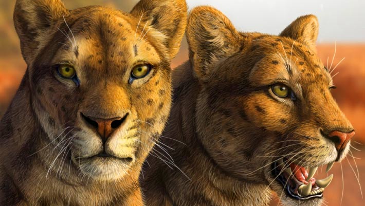
Early Saber-Toothed Cat Gets New Face after Decades of Mystery
tooth paleoart reconstitution United States Pliocene mammals skull
A nearly complete skull unearthed decades ago in Arizona has given paleontologists their clearest look yet at Adelphailurus kansensis, an enigmatic felid species that inhabited North America more than 5 million years ago and occupied an early branch of the saber-toothed cat family tree. The post Early Saber-Toothed Cat Gets New Face after Decades of Mystery appeared first on Sci.News: Breaking Science News.
29/06/2026 sci-news
A Stunning Theropod Skull on Display
museum fossil Tyrannosaurus skull
During a visit to the Museum für Naturkunde (Berlin) we took the opportunity to photograph the beautiful skull of Tristan the Tyrannosaurus rex.  The skull is one of the most complete and best-preserved tyrannosaur skulls known to science.  The T. rex fossils, on display next to the skull material have been named Tristan Otto.  To
12/06/2026 everythingdinosaur
1 2 3 4 5 6