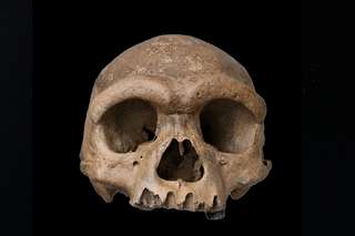
skull

View type

386 image(s) · 29 News

Image gallery

Figure description from paper: "Fig. 3 Wimanius odontopalatus Maisch & Matzke, 1998, holotype, GPIT-PV-76272. Photos courtesy of G. Bindellini (Milano). a The complete holotype. b Detail showing a part of the dentition. c Disarticulated bones from the posterior part of the skull
Taxa Wimaniidae

Figure description from paper: "Fig. 3 Wimanius odontopalatus Maisch & Matzke, 1998, holotype, GPIT-PV-76272. Photos courtesy of G. Bindellini (Milano). a The complete holotype. b Detail showing a part of the dentition. c Disarticulated bones from the posterior part of the skull

bone description holotype Wimaniidae +2
Holotype specimen (PIMUZ A/III 1274) of Prosantosaurus scheffoldi gen. et spec. nov. from the upper Prosanto Formation (Early Ladinian, Middle Triassic) of Ducanfurgga locality no. 4, southwest of Davos, Canton of Grisons, south-eastern Switzerland. A Nearly complete specimen as prepared in dorsal view. The posterior part of the tail was lost prior to burial. Both forearms are not visible but lie below the trunk region, pointing in an anteromedial direction (see Additional file 1: Fig. S3A). B Detail of skull and anterior neck region. C Outline sketch of skull sutures. D Detail of shoulder girdle (claviculae, scapulae) and anterior dorsal vertebrae and ribs. E Detail of right humerus. F Detail of posterior dorsal vertebrae and ribs, sacral vertebrae and ribs, and anterior caudal vertebrae and ribs. G Detail of left ilium and hindlimb. ar articular; as astragalus; bo basioccipital; cal calcaneus; cl clavicula; co coracoid; d dentary; en external naris; eo exoccipital; fe femur; fi fibula; fr frontal; hu humerus; il ilium; in internal naris; is ischium; j jugal; mx maxilla; na naris; o orbit; pa parietal; pl palatine; pmx premaxilla; pof postfrontal; po postorbital; prf prefrontal; pt pterygoid; pu pubis; q quadrate; qj quadratojugal; ti tibia; sacr sacral rib; sc scapula; so supraoccipital; sp splenial; sq squamosal; su surangular; utf upper temporal fenestra; v vomer

Holotype specimen (PIMUZ A/III 1274) of Prosantosaurus scheffoldi gen. et spec. nov. from the upper Prosanto Formation (Early Ladinian, Middle Triassic) of Ducanfurgga locality no. 4, southwest of Davos, Canton of Grisons, south-eastern Switzerland. A Nearly complete specimen as prepared in dorsal view. The posterior part of the tail was lost prior to burial. Both forearms are not visible but lie below the trunk region, pointing in an anteromedial direction (see Additional file 1: Fig. S3A). B Detail of skull and anterior neck region. C Outline sketch of skull sutures. D Detail of shoulder girdle (claviculae, scapulae) and anterior dorsal vertebrae and ribs. E Detail of right humerus. F Detail of posterior dorsal vertebrae and ribs, sacral vertebrae and ribs, and anterior caudal vertebrae and ribs. G Detail of left ilium and hindlimb. ar articular; as astragalus; bo basioccipital; cal calcaneus; cl clavicula; co coracoid; d dentary; en external naris; eo exoccipital; fe femur; fi fibula; fr frontal; hu humerus; il ilium; in internal naris; is ischium; j jugal; mx maxilla; na naris; o orbit; pa parietal; pl palatine; pmx premaxilla; pof postfrontal; po postorbital; prf prefrontal; pt pterygoid; pu pubis; q quadrate; qj quadratojugal; ti tibia; sacr sacral rib; sc scapula; so supraoccipital; sp splenial; sq squamosal; su surangular; utf upper temporal fenestra; v vomer

humerus Switzerland Ladinian Middle Triassic +6
Partial skull and anterior cervical vertebrae of unidentified pliosaurid plesiosaurian (MPPL 18797) from the uppermost Callovian–middle Oxfordian of Kaberlaba (Asiago, Italy), in left lateral view. Photograph (A) and explanatory drawing (B). White, preserved bone surfaces; dark grey, preserved margins of skull fenestrae; light grey, eroded bone surface; cross hatching, broken bone.

Partial skull and anterior cervical vertebrae of unidentified pliosaurid plesiosaurian (MPPL 18797) from the uppermost Callovian–middle Oxfordian of Kaberlaba (Asiago, Italy), in left lateral view. Photograph (A) and explanatory drawing (B). White, preserved bone surfaces; dark grey, preserved margins of skull fenestrae; light grey, eroded bone surface; cross hatching, broken bone.

bone drawing Italy Callovian +6
Holotype of the polycotylid[1] plesiosaur Edgarosaurus muddi DRUCKENMILLER, 2002, (MOR 751) comprising the skull, the neck, and the left front flipper, on display in the Museum of the Rockies in Bozeman, Montana. The specimen was collected in Edgar, Carbon County, Montana, from the Shell Creek member of the Thermopolis Shale Formation, Upper Albian, uppermost Lower Cretaceous.[2]

Holotype of the polycotylid[1] plesiosaur Edgarosaurus muddi DRUCKENMILLER, 2002, (MOR 751) comprising the skull, the neck, and the left front flipper, on display in the Museum of the Rockies in Bozeman, Montana. The specimen was collected in Edgar, Carbon County, Montana, from the Shell Creek member of the Thermopolis Shale Formation, Upper Albian, uppermost Lower Cretaceous.[2]

museum United States Albian Cretaceous +7
Holotype skull of Plesiopleurodon wellesi CM2815 In left and right lateral, dorsal, and oblique left lateral views.

Holotype skull of Plesiopleurodon wellesi CM2815 In left and right lateral, dorsal, and oblique left lateral views.

holotype Plesiopleurodon skull
Skull of the plesiosauroid Lusonectes sauvagei gen et sp. nov. (MG33) from the Toarcian of Portugal, in right lateral (A), left lateral (B), dorsal (C), and ventral (D) views. Photographs (A1–D1), explanatory drawings (A2–D2).

Skull of the plesiosauroid Lusonectes sauvagei gen et sp. nov. (MG33) from the Toarcian of Portugal, in right lateral (A), left lateral (B), dorsal (C), and ventral (D) views. Photographs (A1–D1), explanatory drawings (A2–D2).

drawing Portugal Toarcian Lusonectes +1
Reconstruction of the skull of Lagenanectes richterae in right lateral view. Only the anterior part of the skull and lower jaw is preserved in the type and only known specimen. The posterior parts are reconstructed based on the related genus Libonectes.

Reconstruction of the skull of Lagenanectes richterae in right lateral view. Only the anterior part of the skull and lower jaw is preserved in the type and only known specimen. The posterior parts are reconstructed based on the related genus Libonectes.

specimen Lagenanectes Libonectes skull
A–B, skull in dorsal view; C–E, postcranial skeleton; in left dorsolateral (C) and left lateral (D–E) views. In line drawings (B, E) dark grey tone indicates damage and light grey tone indicates the palate. Abbreviations: ca, caudal vertebra [number following indicates order in preserved series]; ce, cervical vertebra; d, dorsal vertebra; depr, depression; ecto, ectopterygoid; epip, epipterygoid; exp, expanded neural spine apex; fr, frontal; jug, jugal; l., left [followed by name of element]; mx, maxilla; p, ‘pectoral’ vertebra; par, parietal; pmx, premaxilla; po, postorbital; pofr, postfrontal; prfr, prefrontal; qua, quadrate; r., right [followed by name of element]; s, sacral vertebra; sq, squamosal; unexp, unexpanded neural spine apex. Scale bars equal 50 mm (A–B), 20 mm (C), and 200 mm (D–E).

A–B, skull in dorsal view; C–E, postcranial skeleton; in left dorsolateral (C) and left lateral (D–E) views. In line drawings (B, E) dark grey tone indicates damage and light grey tone indicates the palate. Abbreviations: ca, caudal vertebra [number following indicates order in preserved series]; ce, cervical vertebra; d, dorsal vertebra; depr, depression; ecto, ectopterygoid; epip, epipterygoid; exp, expanded neural spine apex; fr, frontal; jug, jugal; l., left [followed by name of element]; mx, maxilla; p, ‘pectoral’ vertebra; par, parietal; pmx, premaxilla; po, postorbital; pofr, postfrontal; prfr, prefrontal; qua, quadrate; r., right [followed by name of element]; s, sacral vertebra; sq, squamosal; unexp, unexpanded neural spine apex. Scale bars equal 50 mm (A–B), 20 mm (C), and 200 mm (D–E).

scale vertebra drawing Avalonnectes +2
Skull of Augustasaurus hagdorni in the Field Museum of Natural History.

Skull of Augustasaurus hagdorni in the Field Museum of Natural History.

museum Augustasaurus skull
A color-coded skull diagram of Palatodonta bleekeri, mostly based on a diagram published by Neenan et al. (2013). Some interpretations (such as the presence of a quadratojugal) are based on Maisch (2020). Depicted in left lateral view, mirrored from the original specimen as preserved in right lateral view. Dotted lines indicate missing bone.

A color-coded skull diagram of Palatodonta bleekeri, mostly based on a diagram published by Neenan et al. (2013). Some interpretations (such as the presence of a quadratojugal) are based on Maisch (2020). Depicted in left lateral view, mirrored from the original specimen as preserved in right lateral view. Dotted lines indicate missing bone.

bone specimen Palatodonta skull
skull restoration of Russellosaurus

skull restoration of Russellosaurus

Russellosaurus skull
Cuspicephalus scarfi MJMG K1918. A, Original skull on slab of mudstone, Lacking the mandible and dentition; B, outline diagram of preserved bone. Light grey is bone, dark grey is fibrous bone of sagittal crest, black is dental alveoli where unambiguous; C restoration of skull outline, with hypothetical lower jaw. Scale bar = 50 mm. Abbreviation: j, jugal; naof, nasoantorbital fenestra; m, maxilla; o, orbit; pm, premaxilla; q, quadrate; sc,

sagittal crest; sq, squamosal; tr np, trace of nasal process.

Cuspicephalus scarfi MJMG K1918. A, Original skull on slab of mudstone, Lacking the mandible and dentition; B, outline diagram of preserved bone. Light grey is bone, dark grey is fibrous bone of sagittal crest, black is dental alveoli where unambiguous; C restoration of skull outline, with hypothetical lower jaw. Scale bar = 50 mm. Abbreviation: j, jugal; naof, nasoantorbital fenestra; m, maxilla; o, orbit; pm, premaxilla; q, quadrate; sc, sagittal crest; sq, squamosal; tr np, trace of nasal process.

bone crest scale Cuspicephalus +1
3D printed skull of Caelestiventus hanseni.

3D printed skull of Caelestiventus hanseni.

Caelestiventus skull
Figure 2.
Holotype of Liaodactylus primus gen. et sp. nov. (PMOL-AP00031): photographs and line drawings of the nearly complete skull with mandibles in left lateral (a,b), and palatal (c,d) views. aa, atlas-axis complex; an, angular; arf, articular facet; bo, basioccipital; bs, basisphenoid; co, coronoid; d, dentary; ec, ectopterygoid; f, frontal; j, jugal; m, maxilla; naof, nasoantorbital fenestra; p, parietal; pal, palatine; pat, proatlas; pecf, pterygo-ectopterygoid fenestra; pm, premaxilla; pof, postfrontal; pra, prearticular; pt, pterygoid; q, quadrate; qj, quadratojugal; sa, surangular; sof, suborbital fenestra; spl, splenial; sq, squamosal; stf, subtemporal fenestra.

Figure 2. Holotype of Liaodactylus primus gen. et sp. nov. (PMOL-AP00031): photographs and line drawings of the nearly complete skull with mandibles in left lateral (a,b), and palatal (c,d) views. aa, atlas-axis complex; an, angular; arf, articular facet; bo, basioccipital; bs, basisphenoid; co, coronoid; d, dentary; ec, ectopterygoid; f, frontal; j, jugal; m, maxilla; naof, nasoantorbital fenestra; p, parietal; pal, palatine; pat, proatlas; pecf, pterygo-ectopterygoid fenestra; pm, premaxilla; pof, postfrontal; pra, prearticular; pt, pterygoid; q, quadrate; qj, quadratojugal; sa, surangular; sof, suborbital fenestra; spl, splenial; sq, squamosal; stf, subtemporal fenestra.

drawing holotype Liaodactylus skull
Holotype skull (IVPP V17083) of Guidraco venator on display at the Paleozoological Museum of China.

Holotype skull (IVPP V17083) of Guidraco venator on display at the Paleozoological Museum of China.

museum China holotype Guidraco +1
Holotype skull and mandible of the anhanguerid Ferrodraco (A), holotype skull and mandible of the anhanguerid Mythunga (B), and holotype mandible of the targaryendraconian Aussiedraco (C).

Holotype skull and mandible of the anhanguerid Ferrodraco (A), holotype skull and mandible of the anhanguerid Mythunga (B), and holotype mandible of the targaryendraconian Aussiedraco (C).

holotype Anhangueridae Aussiedraco Ferrodraco +3
1 2 3 4 5 6 7 8 9 10 11 12 13 14 15 16 17 18 19 20 21 22 23 24 25

News

Les scientifiques pensaient les Denisoviens introuvables : cette découverte spectaculaire change désormais tout
Scientists thought the Denisovans were impossible to find: this spectacular discovery now changes everything
bone DNA China discovery Homo sapiens human origin skull
For fifteen years, the Denisovans had neither face nor outline. Just DNA extracted from a piece of Siberian bone the size of a fingernail. In 2025, an exceptional skull discovered in China changed everything. Homo longi, nicknamed the Dragon Man, finally gives a face to this phantom human group. 
06/06/2026 futura-terre ⚙ Auto-translated
This prehistoric fish may explain how animals first walked on Earth
Antarctica skull
Scientists have peered inside the skull of a 380-million-year-old Antarctic fish that was closely related to the first animals to walk on land, revealing surprising clues about how life began its move out of the water. Using advanced neutron imaging, researchers discovered that Koharalepis jarviki had features suited for living near the water’s surface, including openings in its skull that may have helped it gulp air and a light-sensitive organ linked to day-night rhythms.
25/05/2026 sciencedaily-paleo
T. rex’s tiny arms may have evolved for a surprisingly brutal reason
jaw limb hunting predator prey Dinosauria skull
Why did T. rex have such tiny arms? Scientists now think it’s because its giant head became the ultimate hunting tool. Across multiple dinosaur groups, stronger skulls and crushing jaws evolved alongside shrinking forelimbs, especially in predators hunting enormous prey. In other words, once the bite became deadly enough, the arms may have stopped mattering.
20/05/2026 sciencedaily
Stunning 150-million-year-old stegosaur skull rewrites dinosaur evolution
fossil Dacentrurus Dinosauria Stegosauria discovery evolution skull
A spectacular dinosaur discovery in Spain is giving scientists a rare new look inside the world of stegosaurs. Paleontologists uncovered the best-preserved stegosaur skull ever found in Europe, belonging to the iconic plated dinosaur Dacentrurus armatus, which roamed Earth around 150 million years ago. Because stegosaur skulls are extremely fragile and almost never survive intact, the fossil is helping researchers uncover previously unknown details about how these armored giants evolved.
17/05/2026 sciencedaily
New Triassic Carnivorous Dinosaur Identified from Ghost Ranch
United States Late Triassic Triassic fossil specimen Coelophysis Dinosauria Ptychotherates new species skull
Researchers from Virginia Tech (Virginia, USA) have identified a new species of Late Triassic dinosaur.  The dinosaur has been named Ptychotherates bucculentus.  This new carnivorous dinosaur taxon has been erected based on the detailed examination of an incomplete, well-preserved but jumbled up skull fossil (specimen number CM 31368).  The skull comes from the world-famous Coelophysis
11/05/2026 everythingdinosaur
1 2 3 4 5 6