crâne

Type de vue

386 image(s) · 29 Actualités

Galerie d'images

Reconstruction of the Abelisaurus skull with original bones of the holotype. Museo Provincial Carlos Ameghino, Cipolletti, Argentina. Scale = 10 cm
Taxons Abelisaurus

Reconstruction of the Abelisaurus skull with original bones of the holotype. Museo Provincial Carlos Ameghino, Cipolletti, Argentina. Scale = 10 cm

os écaille Argentine holotype +2
Hualianceratops holotype IVVP V18641, Skull reconstruction
Taxons Hualianceratops

Hualianceratops holotype IVVP V18641, Skull reconstruction

holotype Hualianceratops crâne
Figure 2.
Holotype of Liaodactylus primus gen. et sp. nov. (PMOL-AP00031): photographs and line drawings of the nearly complete skull with mandibles in left lateral (a,b), and palatal (c,d) views. aa, atlas-axis complex; an, angular; arf, articular facet; bo, basioccipital; bs, basisphenoid; co, coronoid; d, dentary; ec, ectopterygoid; f, frontal; j, jugal; m, maxilla; naof, nasoantorbital fenestra; p, parietal; pal, palatine; pat, proatlas; pecf, pterygo-ectopterygoid fenestra; pm, premaxilla; pof, postfrontal; pra, prearticular; pt, pterygoid; q, quadrate; qj, quadratojugal; sa, surangular; sof, suborbital fenestra; spl, splenial; sq, squamosal; stf, subtemporal fenestra.
Taxons Liaodactylus

Figure 2. Holotype of Liaodactylus primus gen. et sp. nov. (PMOL-AP00031): photographs and line drawings of the nearly complete skull with mandibles in left lateral (a,b), and palatal (c,d) views. aa, atlas-axis complex; an, angular; arf, articular facet; bo, basioccipital; bs, basisphenoid; co, coronoid; d, dentary; ec, ectopterygoid; f, frontal; j, jugal; m, maxilla; naof, nasoantorbital fenestra; p, parietal; pal, palatine; pat, proatlas; pecf, pterygo-ectopterygoid fenestra; pm, premaxilla; pof, postfrontal; pra, prearticular; pt, pterygoid; q, quadrate; qj, quadratojugal; sa, surangular; sof, suborbital fenestra; spl, splenial; sq, squamosal; stf, subtemporal fenestra.

dessin holotype Liaodactylus crâne
The skull of Zupaysaurus rougieri (A) in right lateral view. Scale bar equals 5 cm. Cropped from Figure 2 in the source.
anfe - antorbital fenestra
emf - external mandibular fenestra
itf - infratemporal fenestra
or - orbit
Taxons Zupaysaurus

The skull of Zupaysaurus rougieri (A) in right lateral view. Scale bar equals 5 cm. Cropped from Figure 2 in the source. anfe - antorbital fenestra emf - external mandibular fenestra itf - infratemporal fenestra or - orbit

écaille Zupaysaurus crâne
Skeletal elements of Bajadasaurus pronuspinax gen. et sp. nov (MMCh-PV 75). (A–C) Skull roof and braincase in posterior (A), left lateral (B) and right lateral (C) views. (D,E) Left lower jaw in dorsal (D) and medial (E) views. F, Dentaries in anterior view. (G) Pterygoids in ventral view. (H) Left maxilla in medial view. (I) Left lacrimal in lateral view. (J) Left quadratojugal in lateral view. (K,L) Right quadrate in medial (K) and posterior (L) views. (M) Proatlases in dorsal view. (N) Atlantal neurapophyses in anterior view. (O,P), Axis in left lateral (O) and anterior (P) views. (Q,R) Fifth cervical vertebra in left lateral (Q) and anterior (R) views. an, angular; ar, articular; bo, basioccipital; bt, basal tubera; btp, basipterygoid process; ch, ‘chin’ of dentary; cn, cranial nerve; d, dentary; di, diapophysis; f, frontal; fm, foramen magnum; fo, fenestra ovalis; ls, laterosphenoid; met, metotic foramen; mp, medial process; nc, neural canal; ns, neural spine; os, orbitosphenoid; p, parietal; pfo, pneumatic fossa; po, postorbital; pocdf, postzygapophyseal centrodiapophyseal fossa; podl, postzygodiapophyseal lamina; popr, paraoccipital process; poz, postzygapophysis; pra, prearticular; pre, prezygapophysis; prsl, prespinal lamina; ptf, postemporal fenestra; qf, quadrate fossa; rm, replacement maxillary tooth; sa, surangular; sp, splenial; sq, squamosal; stf, supratemporal fenestra; vk, ventral keel; vp, ventral process.
Taxons Bajadasaurus

Skeletal elements of Bajadasaurus pronuspinax gen. et sp. nov (MMCh-PV 75). (A–C) Skull roof and braincase in posterior (A), left lateral (B) and right lateral (C) views. (D,E) Left lower jaw in dorsal (D) and medial (E) views. F, Dentaries in anterior view. (G) Pterygoids in ventral view. (H) Left maxilla in medial view. (I) Left lacrimal in lateral view. (J) Left quadratojugal in lateral view. (K,L) Right quadrate in medial (K) and posterior (L) views. (M) Proatlases in dorsal view. (N) Atlantal neurapophyses in anterior view. (O,P), Axis in left lateral (O) and anterior (P) views. (Q,R) Fifth cervical vertebra in left lateral (Q) and anterior (R) views. an, angular; ar, articular; bo, basioccipital; bt, basal tubera; btp, basipterygoid process; ch, ‘chin’ of dentary; cn, cranial nerve; d, dentary; di, diapophysis; f, frontal; fm, foramen magnum; fo, fenestra ovalis; ls, laterosphenoid; met, metotic foramen; mp, medial process; nc, neural canal; ns, neural spine; os, orbitosphenoid; p, parietal; pfo, pneumatic fossa; po, postorbital; pocdf, postzygapophyseal centrodiapophyseal fossa; podl, postzygodiapophyseal lamina; popr, paraoccipital process; poz, postzygapophysis; pra, prearticular; pre, prezygapophysis; prsl, prespinal lamina; ptf, postemporal fenestra; qf, quadrate fossa; rm, replacement maxillary tooth; sa, surangular; sp, splenial; sq, squamosal; stf, supratemporal fenestra; vk, ventral keel; vp, ventral process.

dent vertèbre Bajadasaurus crâne
The skull of Aquilops in three versions: the actual holotype fossil down below, a reconstruction of the distorted skull with the missing bits sculpted in by preparator and reptile-reconstructor extraordinaire Kyle Davies (see more of his work here), and a reconstruction of the skull as it might have looked before it went through the ravages of taphonomy.
Taxons Aquilops

The skull of Aquilops in three versions: the actual holotype fossil down below, a reconstruction of the distorted skull with the missing bits sculpted in by preparator and reptile-reconstructor extraordinaire Kyle Davies (see more of his work here), and a reconstruction of the skull as it might have looked before it went through the ravages of taphonomy.

fossile holotype Aquilops crâne
Figure 1: Holotype and skeletal reconstruction of Mosaiceratops azumai, gen. et sp. nov (ZMNH M8856). (a) photograph and line drawing of ZMNH M8856; (b) skeletal reconstruction showing preserved elements in white. Scale bar 10 cm. Abbreviations: a, astragalus; boc, basioccipital; c, calcaneum; cav, caudal vertebra; ch, chevron; cv, cervical vertebra; dr, dorsal rib; dv, dorsal vertebra; f, frontal; fem, femur; fl, fibula; h, humerus; il, ilium; is, ischium; L, left; mt, metatarsal; ph, phalanx/phalanges; po, postorbital; R, right; sk, skull; sq, squamosal; t, tibia; td, tendon; ?, undiagnostic remains.
Taxons Mosaiceratops

Figure 1: Holotype and skeletal reconstruction of Mosaiceratops azumai, gen. et sp. nov (ZMNH M8856). (a) photograph and line drawing of ZMNH M8856; (b) skeletal reconstruction showing preserved elements in white. Scale bar 10 cm. Abbreviations: a, astragalus; boc, basioccipital; c, calcaneum; cav, caudal vertebra; ch, chevron; cv, cervical vertebra; dr, dorsal rib; dv, dorsal vertebra; f, frontal; fem, femur; fl, fibula; h, humerus; il, ilium; is, ischium; L, left; mt, metatarsal; ph, phalanx/phalanges; po, postorbital; R, right; sk, skull; sq, squamosal; t, tibia; td, tendon; ?, undiagnostic remains.

humérus écaille vertèbre dessin +3
Skull of Eternauta patagonica (photograph and diagram in right view)
Taxons Eternauta

Skull of Eternauta patagonica (photograph and diagram in right view)

Eternauta crâne
Skeletal diagram featuring the optimal remains of the holotype of Graciliceratops mongoliensis: ZPAL MgD-I/156.[1] Found in the Bayan Shireh Formation, in the original description the remains were referred to the genus Microceratops (now obsolete).[2] However, Sereno in 2000 noted that there was no base for this referral, then, he created a new genus and species for this specimen.[1] The holotype is very fragmented (specially the skull), consisting of:[2]
Fragmented skull; 4 cervical, 12 dorsal and 7 sacral vertebrae; right scapula; proximal end of left scapula; left coracoid; right humerus, radius and fragmentary ulna; proximal and distal end of left humerus; proximal fragments of both pubis; fragments of both illium and fragment of right ischium; right femur, tibia and nearly complete pes; distal part of left tibia, fragmentary left pes; tarsals and isolated ribs.[2]

The sacral vertebrae are not fused, an indicator of the immaturity of this specimen; the estimated adult size is about 2 meters long or similar to Protoceratops.[2][1] Right quadratojugal, quadrate and fragmentary jugal were reversed in order to get an optimal view.
Taxons Graciliceratops

Skeletal diagram featuring the optimal remains of the holotype of Graciliceratops mongoliensis: ZPAL MgD-I/156.[1] Found in the Bayan Shireh Formation, in the original description the remains were referred to the genus Microceratops (now obsolete).[2] However, Sereno in 2000 noted that there was no base for this referral, then, he created a new genus and species for this specimen.[1] The holotype is very fragmented (specially the skull), consisting of:[2] Fragmented skull; 4 cervical, 12 dorsal and 7 sacral vertebrae; right scapula; proximal end of left scapula; left coracoid; right humerus, radius and fragmentary ulna; proximal and distal end of left humerus; proximal fragments of both pubis; fragments of both illium and fragment of right ischium; right femur, tibia and nearly complete pes; distal part of left tibia, fragmentary left pes; tarsals and isolated ribs.[2] The sacral vertebrae are not fused, an indicator of the immaturity of this specimen; the estimated adult size is about 2 meters long or similar to Protoceratops.[2][1] Right quadratojugal, quadrate and fragmentary jugal were reversed in order to get an optimal view.

humérus description holotype spécimen +5
Zhejiangopterus linhaiensis, skull (cast), Zhejiang Geological Museum
Taxons Zhejiangopterus

Zhejiangopterus linhaiensis, skull (cast), Zhejiang Geological Museum

musée moulage Zhejiangopterus crâne
Smitanosaurus agilis skull fragment and neck.
Taxons Smitanosaurus

Smitanosaurus agilis skull fragment and neck.

Smitanosaurus crâne
The theropod skull displays the distinctive features of this apex predator, including a long, robust snout, conical teeth, and strong jaw muscles adapted for gripping and tearing prey.
Taxons Rajasaurus

The theropod skull displays the distinctive features of this apex predator, including a long, robust snout, conical teeth, and strong jaw muscles adapted for gripping and tearing prey.

prédateur proie Rajasaurus crâne
Reconstruction of the skull of Tlatolophus galorum by sculptor Samuel Nieves Tlapaya (El Alebrije Prehistórico). Displayed in the lobby of the hosting hotel of the 12th Latin American Congress of Paleontology, held in Puebla, Mexico, during the first week of March 2025.
Taxons Tlatolophus

Reconstruction of the skull of Tlatolophus galorum by sculptor Samuel Nieves Tlapaya (El Alebrije Prehistórico). Displayed in the lobby of the hosting hotel of the 12th Latin American Congress of Paleontology, held in Puebla, Mexico, during the first week of March 2025.

Mexique Tlatolophus crâne
Skull of Caiuajara dobruskii gen. et sp. nov. (holotype, CP.V 1449) with the shape of an adult individual.

Scale bar equals 50: d, dentary; dcr, dentary crest; dep, depression; exp, ventral expansion of the premaxilla; f, frontal; fcr, frontal crest; fo, foraminae; m, maxilla; oc, occipital condyle; op, opisthotic; p, parietal; pm, premaxilla; pmcr, premaxillary crest; q, quadrate; soc, supraoccipital. The quadrate is inverted.
Taxons Caiuajara

Skull of Caiuajara dobruskii gen. et sp. nov. (holotype, CP.V 1449) with the shape of an adult individual. Scale bar equals 50: d, dentary; dcr, dentary crest; dep, depression; exp, ventral expansion of the premaxilla; f, frontal; fcr, frontal crest; fo, foraminae; m, maxilla; oc, occipital condyle; op, opisthotic; p, parietal; pm, premaxilla; pmcr, premaxillary crest; q, quadrate; soc, supraoccipital. The quadrate is inverted.

crête écaille holotype Caiuajara +1
Minqaria bata skull reconstruction with known elements shown in white.
Taxons Minqaria

Minqaria bata skull reconstruction with known elements shown in white.

Minqaria crâne
Caupedactylus ybaka holotype skull (MN 4726-V) in D, E, left lateral view.
Taxons Caupedactylus

Caupedactylus ybaka holotype skull (MN 4726-V) in D, E, left lateral view.

holotype Caupedactylus crâne
1 2 3 4 5 6 7 8 9 10 11 12 13 14 15 16 17 18 19 20 21 22 23 24 25

Actualités

Les scientifiques pensaient les Denisoviens introuvables : cette découverte spectaculaire change désormais tout
os ADN Chine découverte Homo sapiens origine humaine crâne
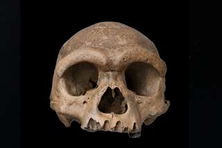
Pendant quinze ans, les Denisoviens n'ont eu ni visage ni contour. Juste de l'ADN extrait d'un fragment d'os sibérien grand comme un ongle. En 2025, un crâne exceptionnel découvert en Chine a tout changé. Homo longi, surnommé l'Homme dragon, donne enfin un visage à ce groupe humain fantôme. 
06/06/2026 futura-terre
Ce poisson préhistorique pourrait expliquer comment les animaux ont marché pour la première fois sur Terre
Antarctique crâne
Les scientifiques ont examiné l'intérieur du crâne d'un poisson de l'Antarctique vieux de 380 millions d'années, étroitement apparenté aux premiers animaux à avoir marché sur terre, révélant des indices surprenants sur la façon dont la vie a commencé à sortir de l'eau. Grâce à l’imagerie neutronique avancée, les chercheurs ont découvert que Koharalepis jarviki possédait des caractéristiques adaptées pour vivre près de la surface de l’eau, notamment des ouvertures dans son crâne qui auraient pu l’aider à avaler de l’air et un organe sensible à la lumière lié aux rythmes jour-nuit.
25/05/2026 sciencedaily-paleo ⚙ Traduction automatique
Les petits bras du T. rex pourraient avoir évolué pour une raison étonnamment brutale
mâchoire membre chasse prédateur proie Dinosauria crâne
Pourquoi le T. rex avait-il des bras si petits ? Les scientifiques pensent désormais que c’est parce que sa tête géante est devenue l’outil de chasse ultime. Dans plusieurs groupes de dinosaures, des crânes plus forts et des mâchoires écrasantes ont évolué parallèlement à des membres antérieurs rétrécis, en particulier chez les prédateurs chassant d'énormes proies. En d’autres termes, une fois que la morsure est devenue suffisamment mortelle, les bras ont peut-être cessé d’avoir de l’importance.
20/05/2026 sciencedaily ⚙ Traduction automatique
Un superbe crâne de stégosaure vieux de 150 millions d'années réécrit l'évolution des dinosaures
fossile Dacentrurus Dinosauria Stegosauria découverte évolution crâne
Une découverte spectaculaire de dinosaures en Espagne donne aux scientifiques un nouveau regard rare sur le monde des stégosaures. Les paléontologues ont découvert le crâne de stégosaure le mieux conservé jamais découvert en Europe, appartenant à l'emblématique dinosaure plaqué Dacentrurus armatus, qui parcourait la Terre il y a environ 150 millions d'années. Parce que les crânes de stégosaures sont extrêmement fragiles et ne survivent presque jamais intacts, le fossile aide les chercheurs à découvrir des détails jusqu'alors inconnus sur l'évolution de ces géants blindés.
17/05/2026 sciencedaily ⚙ Traduction automatique
Un nouveau dinosaure carnivore du Trias identifié à Ghost Ranch
États-Unis Trias supérieur Trias fossile spécimen Coelophysis Dinosauria Ptychotherates nouvelle espèce crâne
Des chercheurs de Virginia Tech (Virginie, États-Unis) ont identifié une nouvelle espèce de dinosaure du Trias supérieur.  Le dinosaure a été nommé Ptychotherates bucculentus.  Ce nouveau taxon de dinosaure carnivore a été érigé sur la base de l'examen détaillé d'un fossile de crâne incomplet, bien conservé mais mélangé (numéro de spécimen CM 31368).  Le crâne provient de la Coelophysis de renommée mondiale
11/05/2026 everythingdinosaur ⚙ Traduction automatique
1 2 3 4 5 6