crâne

Type de vue

386 image(s) · 29 Actualités

Galerie d'images

Identifier: annalsofmedicalh01pack (find matches)
Title: Annals of medical history
Year: 1917 (1910s)
Authors:  Packard, Francis R. (Francis Randolph), 1870-1950
Subjects:  Medicine
Publisher:  New York P.B. Hoeber
Contributing Library:  Gerstein - University of Toronto
Digitizing Sponsor:  University of Toronto

View Book Page: Book Viewer
About This Book: Catalog Entry
View All Images: All Images From Book
Click here to view book online to see this illustration in context in a browseable online version of this book.

Text Appearing Before Image:
ail strongly Hexed, and the toescontracted and appressed. The whole atti-tude strongly suggests a spastic distress,possibly brought on by some form ofpoisoning of the central nervous system,from infection or the deglutition of somepoisonous substance. 14. Osteomalacia is evidently the causeof the hypertrophy of the bones of Lim-nocyron potens, an early carnivore from theWashakie Eocene of Wyoming, nearly3,000,000 years old. MATERIALS AND METHODS The material described in the presentpaper has been loaned the writer lor de-scription by the Field Museum of Chii Osborn: Hull. Amcr. \lus. Natl. Hist., 1917,vol. 35. !>• 733. P>- 28. Studies in Paleopathology 393 by the American Museum of Natural His-tory of New York City, by Walker Museumof the University of Chicago, and by the Uni-versity of Kansas Natural History Museum. made by the well-known petrographic meth-ods so common in all geological labora-tories. The diagnoses, where they are at-tempted, arc made from comparisons of the
Text Appearing After Image:
Fig. 20. The skeleton of Strutbiomimus altus, a small dinosaur from the BeliyRiver series (Cretaceous), Red Deer River, Alberta, Canada, now regarded as ofapproximately the same age as the Judith River series. The unique feature ofthe skull is the total absence of teeth, with a size of skull one-third larger thanthe ostrich and a length of body of about fifteen feet. The position of the skeletonis decidedly that of the opisthotonos which may be regarded as an indication ofdisease. (After Osborn). A beautiful specimen of an osteoma, theonly one known so far, on the vertebra of aKansas Cretaceous mosasaur, was given thewriter by Dr. J. M. Armstrong of St. Paul.The writer expresses his obligations to thegentlemen connected with the above-men-tioned institutions and to Dr. Armstrong.The methods used are a combination ofprocedures in the various lines involved.Microscopic sections, which can be madethin enough for immersion lens study, are material with similar lesions in recent hu-man mat

Note About Images

Please note that these images are extracted from scanned page images that may have been digitally enhanced for readability - coloration and appearance of these illustrations may not perfectly resemble the original work.
Taxons Hexing

Identifier: annalsofmedicalh01pack (find matches) Title: Annals of medical history Year: 1917 (1910s) Authors: Packard, Francis R. (Francis Randolph), 1870-1950 Subjects: Medicine Publisher: New York P.B. Hoeber Contributing Library: Gerstein - University of Toronto Digitizing Sponsor: University of Toronto View Book Page: Book Viewer About This Book: Catalog Entry View All Images: All Images From Book Click here to view book online to see this illustration in context in a browseable online version of this book. Text Appearing Before Image: ail strongly Hexed, and the toescontracted and appressed. The whole atti-tude strongly suggests a spastic distress,possibly brought on by some form ofpoisoning of the central nervous system,from infection or the deglutition of somepoisonous substance. 14. Osteomalacia is evidently the causeof the hypertrophy of the bones of Lim-nocyron potens, an early carnivore from theWashakie Eocene of Wyoming, nearly3,000,000 years old. MATERIALS AND METHODS The material described in the presentpaper has been loaned the writer lor de-scription by the Field Museum of Chii Osborn: Hull. Amcr. \lus. Natl. Hist., 1917,vol. 35. !>• 733. P>- 28. Studies in Paleopathology 393 by the American Museum of Natural His-tory of New York City, by Walker Museumof the University of Chicago, and by the Uni-versity of Kansas Natural History Museum. made by the well-known petrographic meth-ods so common in all geological labora-tories. The diagnoses, where they are at-tempted, arc made from comparisons of the Text Appearing After Image: Fig. 20. The skeleton of Strutbiomimus altus, a small dinosaur from the BeliyRiver series (Cretaceous), Red Deer River, Alberta, Canada, now regarded as ofapproximately the same age as the Judith River series. The unique feature ofthe skull is the total absence of teeth, with a size of skull one-third larger thanthe ostrich and a length of body of about fifteen feet. The position of the skeletonis decidedly that of the opisthotonos which may be regarded as an indication ofdisease. (After Osborn). A beautiful specimen of an osteoma, theonly one known so far, on the vertebra of aKansas Cretaceous mosasaur, was given thewriter by Dr. J. M. Armstrong of St. Paul.The writer expresses his obligations to thegentlemen connected with the above-men-tioned institutions and to Dr. Armstrong.The methods used are a combination ofprocedures in the various lines involved.Microscopic sections, which can be madethin enough for immersion lens study, are material with similar lesions in recent hu-man mat Note About Images Please note that these images are extracted from scanned page images that may have been digitally enhanced for readability - coloration and appearance of these illustrations may not perfectly resemble the original work.

os vertèbre livre musée +11
Holotype of Jaculinykus yaruui gen. et sp. nov. (MPC-D 100/209). (A) Photograph of the specimen. (B) Explanatory drawing of (A). Highlighted areas refer to the indication of the skeletal elements; skull in green, tail in grey, pectoral girdle and forelimbs in red, pelvis and hind limbs in purple. (C) Reconstruction of Jaculinykus yaruui gen. et sp. nov. Grey areas are missing parts.
Taxons Jaculinykus

Holotype of Jaculinykus yaruui gen. et sp. nov. (MPC-D 100/209). (A) Photograph of the specimen. (B) Explanatory drawing of (A). Highlighted areas refer to the indication of the skeletal elements; skull in green, tail in grey, pectoral girdle and forelimbs in red, pelvis and hind limbs in purple. (C) Reconstruction of Jaculinykus yaruui gen. et sp. nov. Grey areas are missing parts.

membre bassin dessin holotype +3
Reconstruction of the skull of Lagenanectes richterae in right lateral view. Only the anterior part of the skull and lower jaw is preserved in the type and only known specimen. The posterior parts are reconstructed based on the related genus Libonectes.
Taxons Lagenanectes

Reconstruction of the skull of Lagenanectes richterae in right lateral view. Only the anterior part of the skull and lower jaw is preserved in the type and only known specimen. The posterior parts are reconstructed based on the related genus Libonectes.

spécimen Lagenanectes Libonectes crâne
Diagram featuring the holotype skull (ZPAL MgD-I/105) of Tylocephale gilmorei in left lateral view. Scale bar equals 5 cm (50 mm). Based on Maryańska & Osmólska, 1974,[1] and Sullivan, 2006.[2]
References

↑ (1974). "Pachycephalosauria, a new suborder of ornithischian dinosaurs". Palaeontologica Polonica (30): 45−102.

↑ (2006). "A taxonomic review of the Pachycephalosauridae (Dinosauria: Ornithischia)". New Mexico Museum of Natural History and Science Bulletin (35): 347–365.
Taxons Tylocephale

Diagram featuring the holotype skull (ZPAL MgD-I/105) of Tylocephale gilmorei in left lateral view. Scale bar equals 5 cm (50 mm). Based on Maryańska & Osmólska, 1974,[1] and Sullivan, 2006.[2] References ↑ (1974). "Pachycephalosauria, a new suborder of ornithischian dinosaurs". Palaeontologica Polonica (30): 45−102. ↑ (2006). "A taxonomic review of the Pachycephalosauridae (Dinosauria: Ornithischia)". New Mexico Museum of Natural History and Science Bulletin (35): 347–365.

écaille musée Mexique holotype +6
Photograph of the holotype skull of Titanoceratops ouranos, from the late Campanian aged Kirtland/Fruitland beds of New Mexico, USA. Photograph by Nick Longrich
Taxons Titanoceratops

Photograph of the holotype skull of Titanoceratops ouranos, from the late Campanian aged Kirtland/Fruitland beds of New Mexico, USA. Photograph by Nick Longrich

Mexique États-Unis Fruitland Kirtland +4
Lateral diagram of the skull of Sinocephale bexelli (Bohlin, 1953) with unknown regions (grey) restored after Stegoceras
Taxons Sinocephale

Lateral diagram of the skull of Sinocephale bexelli (Bohlin, 1953) with unknown regions (grey) restored after Stegoceras

Sinocephale Stegoceras crâne
Cearadactylus atrox holotype (MN 7019-V), A, B, skull in left lateral view; C, skull and lower jaw in right lateral view
Taxons Cearadactylus

Cearadactylus atrox holotype (MN 7019-V), A, B, skull in left lateral view; C, skull and lower jaw in right lateral view

holotype Cearadactylus crâne
Paratype skull of Hamipterus tianshanensis on display at the Paleozoological Museum of China.
Taxons Hamipteridae

Paratype skull of Hamipterus tianshanensis on display at the Paleozoological Museum of China.

musée Chine Hamipteridae Hamipterus +1
Paratype skull of Hamipterus tianshanensis on display at the Paleozoological Museum of China.
Taxons Hamipterus

Paratype skull of Hamipterus tianshanensis on display at the Paleozoological Museum of China.

musée Chine Hamipteridae Hamipterus +1
Reconstruction of the holotype skull (PIN 3906/2) of the Late Cretaceous sauropod Quaesitosaurus orientalis. Based on Kurzanov & Bannikov 1983,[1] missing elements restored after Nemegtosaurus.[2]
Color Key
  Preserved
  Missing
References

↑ (1983). "A new sauropod from the Upper Cretaceous of Mongolia". Paleontological Journal 2: 90−96.

↑ (2005). "Redescription of the mongolian sauropod Nemegtosaurus mongoliensis nowinski (dinosauria: Saurischia) and comments on late cretaceous sauropod diversity". Journal of Systematic Palaeontology 3 (3): 283−318. DOI:10.1017/S1477201905001628.
Taxons Quaesitosaurus

Reconstruction of the holotype skull (PIN 3906/2) of the Late Cretaceous sauropod Quaesitosaurus orientalis. Based on Kurzanov & Bannikov 1983,[1] missing elements restored after Nemegtosaurus.[2] Color Key   Preserved   Missing References ↑ (1983). "A new sauropod from the Upper Cretaceous of Mongolia". Paleontological Journal 2: 90−96. ↑ (2005). "Redescription of the mongolian sauropod Nemegtosaurus mongoliensis nowinski (dinosauria: Saurischia) and comments on late cretaceous sauropod diversity". Journal of Systematic Palaeontology 3 (3): 283−318. DOI:10.1017/S1477201905001628.

Mongolie Crétacé Crétacé supérieur holotype +5
Australian Mesozoic pterosaurs. A, Aussiedraco molnari (QM F10613; holotype) mandible in dorsal view. Scale = 1 cm. B, Mythunga camara (QM F18896; holotype) partial skull and mandible in left lateral view. Scale = 5 cm. C, Thapunngaka shawi (KK F494; holotype) mandible in left lateral view. Scale = 5 cm. D, Ferrodraco lentoni (AODF 0876; holotype [part]) partial skull and mandible in left lateral view. Scale = 5 cm.
Taxons Aussiedraco

Australian Mesozoic pterosaurs. A, Aussiedraco molnari (QM F10613; holotype) mandible in dorsal view. Scale = 1 cm. B, Mythunga camara (QM F18896; holotype) partial skull and mandible in left lateral view. Scale = 5 cm. C, Thapunngaka shawi (KK F494; holotype) mandible in left lateral view. Scale = 5 cm. D, Ferrodraco lentoni (AODF 0876; holotype [part]) partial skull and mandible in left lateral view. Scale = 5 cm.

écaille Mésozoïque holotype Aussiedraco +6
Figure description from paper: "Fig. 3 Wimanius odontopalatus Maisch & Matzke, 1998, holotype, GPIT-PV-76272. Photos courtesy of G. Bindellini (Milano). a The complete holotype. b Detail showing a part of the dentition. c Disarticulated bones from the posterior part of the skull
Taxons Wimanius

Figure description from paper: "Fig. 3 Wimanius odontopalatus Maisch & Matzke, 1998, holotype, GPIT-PV-76272. Photos courtesy of G. Bindellini (Milano). a The complete holotype. b Detail showing a part of the dentition. c Disarticulated bones from the posterior part of the skull

os description holotype Wimanius +1
Holotype skull (IVPP V17083) of Guidraco venator on display at the Paleozoological Museum of China.
Taxons Guidraco

Holotype skull (IVPP V17083) of Guidraco venator on display at the Paleozoological Museum of China.

musée Chine holotype Guidraco +1
Figure 1: Reconstruction of the skull of Bellusaurus sui from the Middle-Late Jurassic Shishugou Formation of Xinjiang, China.
This reconstruction is a composite based on isolated holotypic and referred material. (A) Right lateral view. (B) Dorsal view. Holotypic elements are indicated in blue and referred elements are in green.
Taxons Bellusaurus

Figure 1: Reconstruction of the skull of Bellusaurus sui from the Middle-Late Jurassic Shishugou Formation of Xinjiang, China. This reconstruction is a composite based on isolated holotypic and referred material. (A) Right lateral view. (B) Dorsal view. Holotypic elements are indicated in blue and referred elements are in green.

Chine Jurassique Jurassique supérieur Bellusaurus +2
Photograph of the skulls NHMD 164741 and NHMD 164758. (A) NHMD 164741 in left lateral view. (B) NHMD 164758 in left lateral view.
Taxons Issi

Photograph of the skulls NHMD 164741 and NHMD 164758. (A) NHMD 164741 in left lateral view. (B) NHMD 164758 in left lateral view.

Issi crâne
Known skull material of Kuru kulla (gen. et sp. nov. Napoli et al. 2022) - premaxilla, dentary, surangular, lacrimal, and teeth - shown with a speculative restoration of the remainder of the skull based on the related taxon Velociraptor mongoliensis.  Known skull material is shown in light grey and labeled; unknown parts of the skull are shown in dark grey.
Taxons Kuru

Known skull material of Kuru kulla (gen. et sp. nov. Napoli et al. 2022) - premaxilla, dentary, surangular, lacrimal, and teeth - shown with a speculative restoration of the remainder of the skull based on the related taxon Velociraptor mongoliensis. Known skull material is shown in light grey and labeled; unknown parts of the skull are shown in dark grey.

Kuru Velociraptor crâne
1 2 3 4 5 6 7 8 9 10 11 12 13 14 15 16 17 18 19 20 21 22 23 24 25

Actualités

Les scientifiques pensaient les Denisoviens introuvables : cette découverte spectaculaire change désormais tout
os ADN Chine découverte Homo sapiens origine humaine crâne
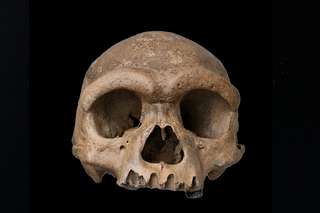
Pendant quinze ans, les Denisoviens n'ont eu ni visage ni contour. Juste de l'ADN extrait d'un fragment d'os sibérien grand comme un ongle. En 2025, un crâne exceptionnel découvert en Chine a tout changé. Homo longi, surnommé l'Homme dragon, donne enfin un visage à ce groupe humain fantôme. 
06/06/2026 futura-terre
Ce poisson préhistorique pourrait expliquer comment les animaux ont marché pour la première fois sur Terre
Antarctique crâne
Les scientifiques ont examiné l'intérieur du crâne d'un poisson de l'Antarctique vieux de 380 millions d'années, étroitement apparenté aux premiers animaux à avoir marché sur terre, révélant des indices surprenants sur la façon dont la vie a commencé à sortir de l'eau. Grâce à l’imagerie neutronique avancée, les chercheurs ont découvert que Koharalepis jarviki possédait des caractéristiques adaptées pour vivre près de la surface de l’eau, notamment des ouvertures dans son crâne qui auraient pu l’aider à avaler de l’air et un organe sensible à la lumière lié aux rythmes jour-nuit.
25/05/2026 sciencedaily-paleo ⚙ Traduction automatique
Les petits bras du T. rex pourraient avoir évolué pour une raison étonnamment brutale
mâchoire membre chasse prédateur proie Dinosauria crâne
Pourquoi le T. rex avait-il des bras si petits ? Les scientifiques pensent désormais que c’est parce que sa tête géante est devenue l’outil de chasse ultime. Dans plusieurs groupes de dinosaures, des crânes plus forts et des mâchoires écrasantes ont évolué parallèlement à des membres antérieurs rétrécis, en particulier chez les prédateurs chassant d'énormes proies. En d’autres termes, une fois que la morsure est devenue suffisamment mortelle, les bras ont peut-être cessé d’avoir de l’importance.
20/05/2026 sciencedaily ⚙ Traduction automatique
Un superbe crâne de stégosaure vieux de 150 millions d'années réécrit l'évolution des dinosaures
fossile Dacentrurus Dinosauria Stegosauria découverte évolution crâne
Une découverte spectaculaire de dinosaures en Espagne donne aux scientifiques un nouveau regard rare sur le monde des stégosaures. Les paléontologues ont découvert le crâne de stégosaure le mieux conservé jamais découvert en Europe, appartenant à l'emblématique dinosaure plaqué Dacentrurus armatus, qui parcourait la Terre il y a environ 150 millions d'années. Parce que les crânes de stégosaures sont extrêmement fragiles et ne survivent presque jamais intacts, le fossile aide les chercheurs à découvrir des détails jusqu'alors inconnus sur l'évolution de ces géants blindés.
17/05/2026 sciencedaily ⚙ Traduction automatique
Un nouveau dinosaure carnivore du Trias identifié à Ghost Ranch
États-Unis Trias supérieur Trias fossile spécimen Coelophysis Dinosauria Ptychotherates nouvelle espèce crâne
Des chercheurs de Virginia Tech (Virginie, États-Unis) ont identifié une nouvelle espèce de dinosaure du Trias supérieur.  Le dinosaure a été nommé Ptychotherates bucculentus.  Ce nouveau taxon de dinosaure carnivore a été érigé sur la base de l'examen détaillé d'un fossile de crâne incomplet, bien conservé mais mélangé (numéro de spécimen CM 31368).  Le crâne provient de la Coelophysis de renommée mondiale
11/05/2026 everythingdinosaur ⚙ Traduction automatique
1 2 3 4 5 6