Galerie d'images
Toutes les images de la base — taxons, formations et intervalles géologiques.
⚠ La fonctionnalité de récupération des images est en cours de test, des images non pertinentes peuvent apparaître.
4,826 image(s)

Mandibular symphyses of Xericeps curvirostris. A–E Holotype FSAC-KK 10700; F–H referred specimen FSAC-KK 5203. A, F In left lateral view; B, G in occlusal view; C, H in ventral view; D, in posterior view and E, in anterior view. Scale bars represent 10 mm. A-C Ammonium chloride coated specimen

Photographs of the holotype of Europejara olcadesorum gen. et sp. nov. (MCCM-LH 9413). (A) Main slab under ultraviolet light. (B) Acid-prepared counterslab. Scale bar: 50 mm.


Nemicolopterus crypticus holotype (IVPP V-14377). (A) Skeleton overview, and (B) schematic drawing. (C) Skull (right lateral view), and (D) schematic drawing. Abbreviations: cdv, caudal vertebrae; co, coracoid; cv, cervical vertebra; d1–d4, digits 1–4; f, frontal; fe, femur; h, humerus; hy, hyoid; il, illium; is, ischium; j, jugal; l, left; la, lacrimal; mand, mandible; mc, metacarpal; mt, metatarsal; naof, nasoantorbital fenestra; or, orbit; pa, parietal; pm, premaxilla; ph, phalanx; ti, tibia; r, right; sca, scapula. Scale bars: A–B, 100 mm; C–D, 5 mm.


Aurorazhdarcho micronyx (specimen formerly classified as Pterodactylus longirostris) in Exposition temporaire: Perles

Partial wing of a czech pterosaur Cretornis hlavaci, discovered in 1880 near Choceň. National Museum in Prague.

CAMSM B.54485, holotype, rostrum fragment of Ornithostoma sedgwicki, in posterior, lateral, anterior, and ventral views. Owen's original, inccurate caption: Fig. 4. Side view of part of the proximal end of the metacarpal of the 5th or wing-finger. Fig. 5. Back (?) view of part of the proximal end of the metacarpal of the 5th or wing-finger.


Holotype rostrum FSAC-KK 5075 (A–E) and paratype mandibular symphysis FSAC-KK 5076 (F–H) of Leptostomia begaaensis. A in dorsal view; B, G in occlusal view; C, H in right lateral view; D in anterior view; E in posterior view and F in ventral view. Scale bars A–D, F–H represent 5 mm; D-E 1 mm

Restoration of Domeykodactylus ceciliae based on images of known remains and related genera.

Holotype jaw fragment of Banguela oberlii (or Thalassodromeus, inaccurately referred to as T. sethi in source[1]) with cross-section (line of section indicated by red line).

Partial rostrum of Iberodactylus andreui gen. et sp. nov. (MPZ-2014/1). Photographic images (A–F) and 3D renderings obtained from μCT data (G–L) in right lateral (A,G), left lateral (B,H), dorsal (C,I), palatal (D,J), caudal (E,K), and cranial (F,L) views. Scale bar in cm. Abbreviations. pm: premaxilla; pmcr: premaxillary crest; prid: palatal ridge; te: teeth; trb: trabeculae.

Australian Mesozoic pterosaurs. A, Aussiedraco molnari (QM F10613; holotype) mandible in dorsal view. Scale = 1 cm. B, Mythunga camara (QM F18896; holotype) partial skull and mandible in left lateral view. Scale = 5 cm. C, Thapunngaka shawi (KK F494; holotype) mandible in left lateral view. Scale = 5 cm. D, Ferrodraco lentoni (AODF 0876; holotype [part]) partial skull and mandible in left lateral view. Scale = 5 cm.


Selected elements used in the diagnosis of Saltriovenator zanellai n. gen. n. sp. Right humerus in medial (A), frontal (B) and distal (C) views; (D) left scapula, medial view; (E) right scapular glenoid and coracoid, lateral view; (F) furcula, ventral view; tooth, labial (G) and apical (H) views; (I) left humerus, medial view; right second metacarpal in dorsal (J), lateral (L) and distal (N) views; first phalanx of the right second digit in dorsal (K), lateral (M) and proximal (O) views; (P–T) right third digit in proximal, dorsal and lateral views; (U) right distal tarsal IV, proximal view; third right metatarsal in proximal (V) and frontal (X) views; second right metatarsal, proximal (W) and frontal (Y) views; (Z) reconstructed skeleton showing identified elements (red). Abbreviations as in text, asterisks mark autapomorphic traits. Scale bars: 10 cm in (A)–(E), (I), and (U)–(Y); two cm in (F), and (J)–(T); one cm in (G).


Skeletal diagram Fosterovenator based on the holotype, silhouette based in skeletal diagram of Ceratosaurus by Scott Hartman.


Holotype right pes of Vespersaurus paranaensis gen. et sp. nov. (MPCO.V 0065d1). (a) Distal end of metatarsal II-IV in dorsal view. (b) Distal part of digit II in lateral view. (c) Distal part of digit IV in lateral view. (d) Entire foot as preserved in lateral view. Anatomical abbreviations: ed, extensor depression; fd, flexor depression; lcp, lateral collateral pit; mc, lateral crest; plg, proximolateral groove; vg, vertical groove; vlg, ventral lateral groove; vm, ventral margin.

Life restoration of Huinculsaurus based in the skeletal diagram of Elaphrosaurus by GetAwayTrike.

A Berthasaura leopoldinae 3d Reconstruction by Alex Pritchard Dinosauria - Dinosaur Fossil Replicas


Porción anterior del cráneo del dinosaurio terópodo Genyodectes saurus. Se observan sus poderosos y afilados dientes, típicos de las formas carnívoras. Museo de Ciencias Naturales de La Plata.

Taken at the National Geographic Museum Spinosaurus Exhibit. Photo by www.ieonexus.com


Fossil of Tarascosaurus salluvicus, an extinct theropod discovered in the South of France
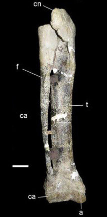
Articulated tibia, fibula, astragalus and calcaneum of Xenotarsosaurus (UNPSJB PV 194/1; Universidad Nacional de la Patagonia ‘San Juan Bosco’, Chubut, Argentina). Scale bar: 5 cm.


Skeletal reconstruction of the Spectrovenator ragei holotype (MZSP-PV 833). Recovered bones in blue.

Abelisaurid theropod Kryptops palaios gen. et sp. nov. MNN GAD1−1 from the Lower Cretaceous Elrhaz Formation of Niger. Left maxilla in lateral view; stereopair (A) and line drawing (B). Cross−hatching indicates broken bone; dashed lines indicate estimated edges.

The tibia of Lametasaurus indicus in different views. The length of the tibia is said to be 58 cm


Fig. 2. Right tibia of Quilmesaurus curriei compared to other tibiae of Abelisauridae. A-B: Quilmesaurus curriei in lateral and anterior view; C: Aucasaurus garridoi, D: Genusaurus sisterornis, E and H: Ekrixinatosaurus novasi, F: Indosuchus raptorius (Courtesy of Dr. F. E. Novas), G: Rajasaurus narmadensis; I: Pycnonemosaurus nevesi Abbreviations: faap: facet for the ascending process of the astragalus; cc: cnemial crest; lm: lateral malleolus; lma: lateral margin of the malleolus; lpcc: lateral process of the cnemial crest; mm: medial malleolus; mma: medial margin of the malleolus; vmcc: ventral margin of the cnemial crest; vpcc: ventral process of the cnemial crest.
Fig. 2. Right tibia of Quilmesaurus curriei compared to other tibiae of Abelisauridae. A-B: Quilmesaurus curriei in lateral and anterior view; C: Aucasaurus garridoi, D: Genusaurus sisterornis, E and H: Ekrixinatosaurus novasi, F: Indosuchus raptorius (Courtesy of Dr. F. E. Novas), G: Rajasaurus narmadensis; I: Pycnonemosaurus nevesi Abbreviations: faap: facet for the ascending process of the astragalus; cc: cnemial crest; lm: lateral malleolus; lma: lateral margin of the malleolus; lpcc: lateral process of the cnemial crest; mm: medial malleolus; mma: medial margin of the malleolus; vmcc: ventral margin of the cnemial crest; vpcc: ventral process of the cnemial crest.

Carnotaurus in Chlupáč museum in Prague
Carnotaurus in Chlupáč museum in Prague

Hypothetical reconstruction of the Late Cretaceous abelisaurid Elemgasem nubilus.


The theropod skull displays the distinctive features of this apex predator, including a long, robust snout, conical teeth, and strong jaw muscles adapted for gripping and tearing prey.


Reconstructed skull of Duriavenator hesperis showing known material (white) of the holotype and only known specimen. Unknown bones based on related Eustreptospondylus oxoniensis. Scale bar is 10cm, image is 10px/cm. Cranial anatomy based on Benson (2008) "A redescription of "Megalosaurus" hesperis (Dinosauria, Theropoda) from the Inferior Oolite (Bajocian, Middle Jurassic) of Dorset, United Kingdom"